
Endoscopic system and evaluation value calculation device

A technology for endoscopy and evaluation results, applied to endoscopy, computing, closed-circuit television systems, etc., can solve problems such as inability to diagnose lesion, achieve the effect of suppressing processing load and suppressing fluctuations

Active Publication Date: 2017-08-18
HOYA CORP
View PDF8 Cites 5 Cited by
  • Summary
  • Abstract
  • Description
  • Claims
  • Application Information

AI Technical Summary

Problems solved by technology

In addition, even skilled operators cannot easily and confidently diagnose a lesion based on subtle differences in color, requiring careful work.

Method used


Image

  • Endoscopic system and evaluation value calculation device
  • Endoscopic system and evaluation value calculation device
  • Endoscopic system and evaluation value calculation device

Examples


Embodiment Construction

[0055] Hereinafter, embodiments of the present invention will be described with reference to the drawings. In addition, in the following description, as one embodiment of the present invention, an electronic endoscope system will be described as an example.

[0056] [Configuration of Electronic Endoscope System 1]

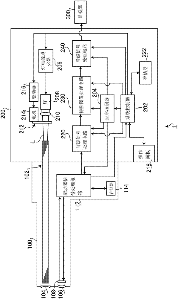
[0057] figure 1 It is a block diagram showing the configuration of the electronic endoscope system 1 according to one embodiment of the present invention. Such as figure 1 As shown, the electronic endoscope system 1 is a system specially used for medical treatment, including an electronic endoscope 100 , a processor 200 and a monitor 300 . The electronic mirror 100 has an insertion portion, and the insertion portion has a front end portion and a curved portion. An LCB (Light Carrying Bundle) 102 is extended in the insertion portion, and a light distribution lens 104 , an objective lens 106 , a solid-state imaging element 108 , and the like are arranged at the f...


PUM


Abstract

This endoscopic system is configured to be provided with: a light source device for irradiating an illumination light toward a subject; an image acquisition unit for capturing reflected light from the subject by an imaging element and acquiring a color image including at least three color components; and an evaluation unit that, inside a color plane defined by at least two color components of the at least three color components, works out evaluation results relating to a target disease of each pixel on the basis of an angle formed by line segments connecting a prescribed reference point set inside the color plane and points corresponding to pixels in the color plane for each pixel constituting the color image acquired by the image acquisition unit, and a reference axis correlated with the target disease. The reference axis is set so as to pass through the prescribed reference point.

Description

technical field [0001] The present invention relates to an endoscope system and an evaluation value calculation device for calculating predetermined evaluation values. Background technique [0002] In general, the lesion will show a different color from the normal mucosal tissue. In recent years, with the improvement of the performance of color endoscope devices, operators have been able to reliably diagnose lesions that are slightly different in color from normal tissues. However, in order for an operator to correctly and reliably diagnose a lesion from a normal tissue based on subtle differences in color on an image captured by an endoscope, long-term training is required under the guidance of skilled technicians. Furthermore, even a skilled operator cannot easily and reliably diagnose a lesion based on a slight difference in color, and careful work is required. [0003] Therefore, for example, Japanese Unexamined Patent Application Publication No. 2014-18332 (hereinafte...

Claims


Application Information

Patent Timeline
Patent Type & Authority Applications(China)
IPC IPC(8): A61B1/04A61B1/00G02B23/24G06T1/00H04N7/18
CPCG02B23/2469H04N7/183G06T7/0012G06T2207/10024G06T2207/10068G06T2207/30096G06T7/80A61B1/00186A61B1/05A61B1/0669A61B1/07A61B1/00009A61B1/00045A61B1/045A61B1/0684A61B1/0655A61B1/0646
Inventor 牧野贵雄
Owner HOYA CORP
Learn More