
A super-resolution microscopic imaging device and method for a micro-droplet lens

A microscopic imaging and super-resolution technology, applied in the field of biomedical detection, can solve the problems of limited microscope popularization, complex focusing, and large size, and achieve the effects of fast enhanced display, broad prospects, and universal practical value

Active Publication Date: 2019-08-13
DALIAN UNIV OF TECH
View PDF1 Cites 0 Cited by
  • Summary
  • Abstract
  • Description
  • Claims
  • Application Information

AI Technical Summary

Problems solved by technology

[0004] Optical microscopes are widely used in biomedical inspections, but their large size, high cost, and complex focusing limit their applications in many fields and regions. The expensive lens group especially limits the popularity of microscopes, and due to the existence of the diffraction limit, The resolution of traditional optical microscopes can actually only reach about 300nm
The existence of the diffraction limit is due to the loss of evanescent waves in the far field

Method used


Image

  • A super-resolution microscopic imaging device and method for a micro-droplet lens

Examples


Embodiment Construction

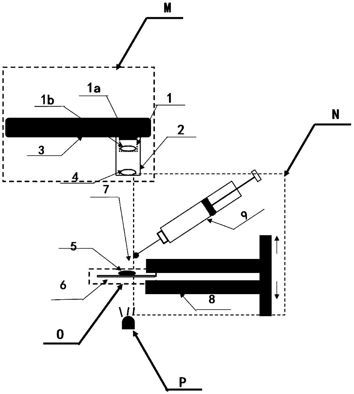
[0010] figure 1 A schematic structural diagram of the device of the present invention is shown, and the smart phone 3 may be a mobile device with an Android, ios or windows phone operating system and a built-in camera module. Wherein the microscope sleeve is configured to accommodate the smart phone camera 1 , the lens 4 , the micro-droplet 7 and the sample 5 to be tested in the same optical path in a way that the illumination path of the illumination light source P is aligned.

[0011] When performing microscopic imaging, the prepared sample 5 to be tested is placed on the slide glass 6, the prepared diesel oil emulsion is injected into the liquid phase micro-syringe 9, and a small amount of micro-droplets 7 are dropped on the sample to be observed. 5 or more. The slide glass is clamped in the precision focusing translation stage 8 . The translation stage can be adjusted up and down to adjust the focal length until a clearly visible image appears on the microscope observati...


PUM


Abstract

The invention relates to a super-resolution micro-imaging device and method of a micro-droplet lens and the device and method can be applied to the fields of water quality environmental monitoring and biomedical diagnosis. With a smart phone or an ordinary personal computer as an observation platform, an optical microscope and a liquid-phase microsyringe are combined; and the liquid-phase microsyringe is used for adjusting the size of the micro-droplet lens for magnifying and displaying a to-be-tested sample so as to adjust a magnification factor of the micro lens, thereby realizing an effect of resolution improvement. Besides, on the basis of combination of a need of an observer, an algorithm for enhanced displaying of an image is embedded into the observation platform, so that various application environment requirements can be met. The system has the good biomedicine detection application prospects. And the super-resolution micro-imaging device and method can be applied to exceed the diffraction limit of the traditional microscope under the optical microscope platform.

Description

[0001] manual [0002] The invention belongs to the field of biomedical detection, and relates to a super-resolution microscopic imaging device and method of a micro-droplet lens, and is a super-resolution microscopic imaging device and method based on a micro-droplet lens with adjustable magnification. technical field [0003] The present invention relates to the field of biomedical detection, and more specifically to a super-resolution microscopic imaging device and method based on a micro-droplet lens with adjustable magnification, which is used to overcome the optical diffraction limit and perform super-resolution microscopic imaging. Background technique [0004] Optical microscopes are widely used in biomedical inspections, but their large size, high cost, and complex focusing limit their applications in many fields and regions. The expensive lens group especially limits the popularity of microscopes, and due to the existence of the diffraction limit, The resolution of...

Claims


Application Information

Patent Timeline
Patent Type & Authority Patents(China)
IPC IPC(8): G02B21/00G02B21/06G02B21/36G01N21/01
CPCG01N21/01G02B21/0004G02B21/06G02B21/36
Inventor 王伟明黄辉喻言欧进萍
Owner DALIAN UNIV OF TECH
Learn More