Human intrahepatic cholangiocarcinoma cell line with high tumorigenic ability and application thereof
A technique for intrahepatic cholangiocarcinoma and cholangiocarcinoma, which is applied in the field of oncology and cell culture, and can solve the problems of unsatisfactory line establishment, unfavorable in vivo experiments, difficult tumor cell models, etc.
- Summary
- Abstract
- Description
- Claims
- Application Information
AI Technical Summary
Problems solved by technology
Method used
Image


Examples
Embodiment 1
[0055] Example 1. Obtainment and observation of human intrahepatic cholangiocarcinoma cell line IHC-ST1
[0056] 1. Primary culture
[0057] The IHC-ST1 cells of the present invention come from the liver tumor tissue in situ of a female patient with intrahepatic cholangiocarcinoma. Under sterile conditions, the liver tumor tissue in situ from a female patient undergoing radical resection of intrahepatic cholangiocarcinoma was taken and rinsed in D-Hanks solution (containing 1000 units / ml penicillin, 3 μg / ml amphotericin B) to remove blood stains and necrotic tissue. Then place the tissue in serum-free DMEM / F12 medium and cut it into 1-3mm with ophthalmic scissors 3 Then add 0.5mg / ml type I collagenase and digest at 37°C for 30min. Fetal calf serum was added to stop the digestion, and the tissue block was filtered through a 200-mesh cell sieve along with the soaking solution, and the filtrate was transferred to a centrifuge tube, and centrifuged at 1000 rpm for 5 minutes. T...
Embodiment 2
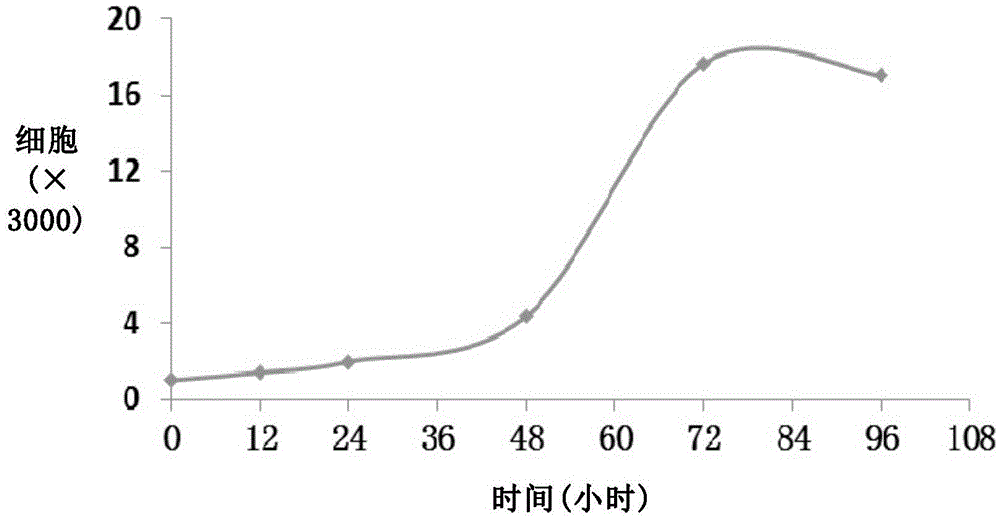
[0065] Example 2, the growth curve of the human intrahepatic cholangiocarcinoma cell line IHC-ST1 of the present invention
[0066] The human intrahepatic cholangiocarcinoma cell line IHC-ST1 cells in good growth state were taken, digested, made into single cell suspension, and counted. Cells were seeded into 96-well plates with 3000 cells per well. CCK-8 detection was carried out at 12h, 24h, 48h, and 72h after cell inoculation, and the average number and standard deviation of the cell number were calculated, and the cell growth curve was drawn.
[0067] The growth curve obtained is as figure 2 . It can be seen from the figure that the cells grow logarithmically on the 1st to 3rd day, and reach a plateau after the 3rd day, and the doubling time is 24h.
Embodiment 3
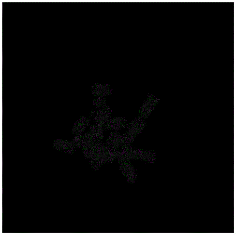
[0068] Example 3. Karyotype analysis of the human intrahepatic cholangiocarcinoma cell line IHC-ST1 of the present invention
[0069] Cells in exponential growth phase, 80%-90% confluent monolayer culture were taken. Add colchicine to suppress metaphase division, so that the final concentration in the medium is 0.04-0.1μg / ml medium, CO 2 Continue culturing for 4 hours in the incubator.
[0070] After fixed staining, the chromosomes of 30 cells in metaphase were counted. The number of chromosomes in the IHC-ST1 cell line was mainly concentrated in 55-70, and the chromosome mode was 62, suggesting the existence of hyperdiploidy. See the representative picture image 3 .
PUM
 Abstract
Description
Claims
Application Information
 - R&D
- Intellectual Property
- Life Sciences
- Materials
- Tech Scout
- Unparalleled Data Quality
- Higher Quality Content
- 60% Fewer Hallucinations
Browse by: Latest US Patents, China's latest patents, Technical Efficacy Thesaurus, Application Domain, Technology Topic, Popular Technical Reports.
© 2025 PatSnap. All rights reserved.Legal|Privacy policy|Modern Slavery Act Transparency Statement|Sitemap|About US| Contact US: help@patsnap.com