Venous blood sampling needle inserting point automatic extraction method based on vision

An automatic extraction and vein technology, applied in image analysis, image enhancement, instruments, etc., can solve the problems of difficult blood collection operation, high risk, high risk coefficient, etc., to facilitate popularization and application, improve efficiency and accuracy, and high flexibility Effect

Active Publication Date: 2017-09-22
XIEHE HOSPITAL ATTACHED TO TONGJI MEDICAL COLLEGE HUAZHONG SCI & TECH UNIV +1
View PDF6 Cites 5 Cited by
  • Summary
  • Abstract
  • Description
  • Claims
  • Application Information

AI Technical Summary

Problems solved by technology

[0002] Medical personnel will frequently collect blood samples during physical examination and diagnosis of patients or during daily inspections. At present, most blood collection occasions use traditional blood collection methods, which require manual operation.
This artificial blood collection method is inefficient and often cannot meet the blood collection requirements during peak hours, and because the blood collection operation is difficult and the risk factor is high, it requires high skills for medical staff, and some patients even have thicker fat and darker skin. Blood vessels cannot be clearly displayed due to other reasons, which leads to puncture failure and brings a lot of inconvenience to medical staff and patients
[0003] Aiming at the difficulty, high risk, and low efficiency of manual blood collection, relevant technical personnel in the field have done some research. For exam

Method used


Image

  • Venous blood sampling needle inserting point automatic extraction method based on vision
  • Venous blood sampling needle inserting point automatic extraction method based on vision

Examples


Embodiment Construction

[0026] In order to make the object, technical solution and advantages of the present invention clearer, the present invention will be further described in detail below in conjunction with the accompanying drawings and embodiments. It should be understood that the specific embodiments described here are only used to explain the present invention, not to limit the present invention. In addition, the technical features involved in the various embodiments of the present invention described below can be combined with each other as long as they do not constitute a conflict with each other.

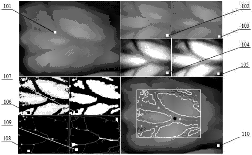
[0027] see figure 1 and figure 2 According to the vision-based automatic extraction method of puncture points for venous blood collection provided by a preferred embodiment of the present invention, the automatic extraction method for puncture points of venous blood collection mainly includes the following steps:

[0028] Step 1: Take an infrared image of the body part where the puncture poin...


PUM


Abstract

The invention belongs to the technical field of machine vision and medical instruments and discloses a venous blood sampling needle inserting point automatic extraction method based on vision. The method comprises the following steps of carrying out whole skeleton extraction on venous bifurcations in a binary image, thereby obtaining an image containing a venous bifurcation center line, further obtaining intersection points of the venous bifurcations, and moreover, recording the coordinates of the intersection points of the venous bifurcations; carrying out partial skeleton extraction on the venous bifurcations in the binary image, thereby obtaining a partially refined image; processing the partially refined image according to the obtained coordinates of the intersection points of the venous bifurcations, carrying out statistics on the number of valid points of each intersection point in a preset length range of the midpoint of the intersection point column by column from the venous proximal part corresponding to the orientation of the intersection point, and further obtaining a length count value corresponding to each intersection point, wherein the interaction point corresponding to the venous bifurcation with the maximum length count value is the venous blood sampling needle inserting point. According to the method, the needle inserting point is extracted automatically and the precision and efficiency are improved.

Description

technical field [0001] The invention belongs to the related technical fields of machine vision and medical instruments, and more specifically relates to a vision-based automatic extraction method for venous blood collection needle points. Background technique [0002] Medical staff will frequently collect blood samples during physical examination and diagnosis of patients or during daily inspections. At present, most blood collection occasions use traditional blood collection methods, which require manual operations. This artificial blood collection method is inefficient and often cannot meet the blood collection requirements during peak hours, and because the blood collection operation is difficult and the risk factor is high, it requires high skills for medical staff, and some patients even have thicker fat and darker skin. And other reasons lead to blood vessels can not be clearly presented, and then lead to puncture failure, bring a lot of inconvenience to medical staff ...

Claims


Application Information

Patent Timeline
IPC IPC(8): G06T7/00G06T7/11
CPCG06T7/0012G06T7/11G06T2207/10048G06T2207/30101G06T2207/30172
Inventor 王国斌陈学东叶霖韩斌郭晓阳皮堂正
Owner XIEHE HOSPITAL ATTACHED TO TONGJI MEDICAL COLLEGE HUAZHONG SCI & TECH UNIV
Why Eureka
Try Eureka