Method and device for extracting pulmonary lobe from chest CT image

A CT image and lung lobe technology, applied in the computer field, can solve the problems of quantitative evaluation of lung lobes, the inability to quantitatively evaluate the condition of a single lung lobe, and affecting the treatment effect of COPD.

Active Publication Date: 2017-10-03
NORTHEASTERN UNIV
View PDF6 Cites 38 Cited by
  • Summary
  • Abstract
  • Description
  • Claims
  • Application Information

AI Technical Summary

Problems solved by technology

[0003] Because CT equipment can obtain clear chest CT images, the use of CT images has become a major means of diagnosing COPD. However, the current medical level can only achieve quantitative diagnosis of a single lung. Evaluation c

Method used


Image

  • Method and device for extracting pulmonary lobe from chest CT image
  • Method and device for extracting pulmonary lobe from chest CT image
  • Method and device for extracting pulmonary lobe from chest CT image

Examples


Embodiment Construction

[0086] In order to better explain the present invention and facilitate understanding, the following describes the present invention in detail through specific embodiments in conjunction with the accompanying drawings.

[0087] Preferred embodiment

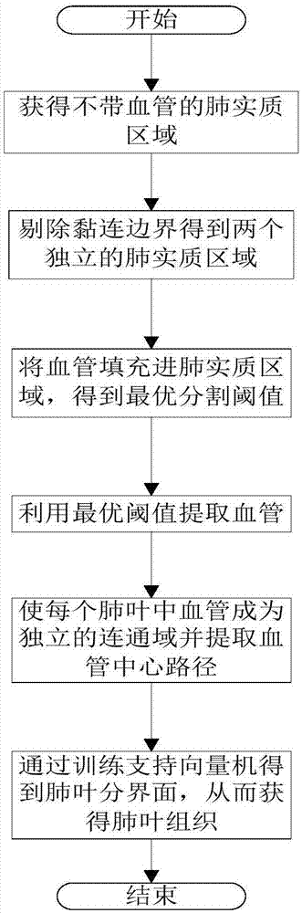
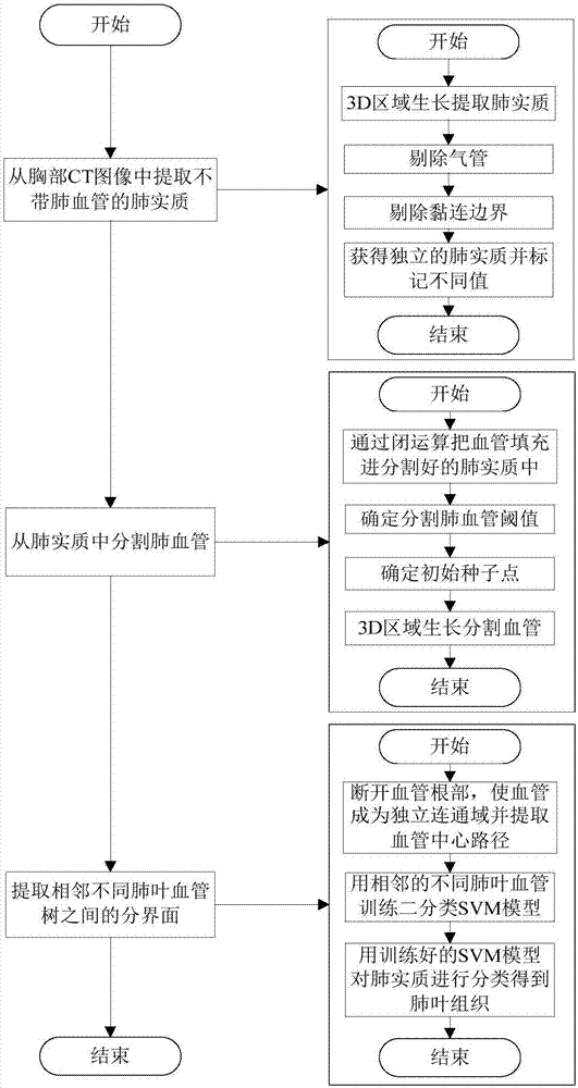
[0088] Such as figure 1 with figure 2 As shown, this embodiment proposes a preferred method for extracting lung lobes from a chest CT image. The method for extracting lung lobes from a chest CT image includes the following steps:

[0089] S1. Receive the input n (n is a natural number) chest CT image, obtain the intermediate image layer, select the specified pixel point of the lung area as the seed point, and perform 3D area growth according to the set segmentation threshold and the initial seed point to obtain the without Pulmonary parenchymal area of ​​pulmonary blood vessels.

[0090] Specifically, step S1 includes the following steps:

[0091] S11. Receive the input n (n is a natural number) chest CT image, obtain an intermediate image...


PUM


Abstract

The invention discloses a method and device for extracting a pulmonary lobe from a chest CT image and relates to the technical field of a computer. The method can obtain pulmonary lobe tissues through a pulmonary parenchyma extraction process based on 3D region growth, a left and right lung adhesion trachea removal process based on region features, a pulmonary vascular root removal process, a pulmonary vascular center path extraction process based on topological thinning and a pulmonary lobe segmentation algorithm based on support vector classification. The method can accurately extract the pulmonary lobe from the chest CT image, can accurately finish quantitative evaluation of illness degree of each pulmonary lobe and is more accurate and effective for diagnosis and treatment of lung diseases.

Description

Technical field [0001] The invention belongs to the field of computer technology and relates to a method and device for extracting lung lobes from chest CT images. Background technique [0002] CT (Computed Tomography) uses a precisely collimated X-ray beam and a highly sensitive detector to scan a certain part of the human body one by one. Different gray levels indicate the degree of X-ray absorption by organs and tissues. For example, on a chest CT image, low-density areas indicate trachea and lung parenchyma, and high-density areas indicate blood vessels, thoracic cavity, bones, etc., which have the characteristics of fast scanning time and clear images, which can be used for inspection of various diseases. Doctors observe and diagnose diseases to provide a convenient and reliable basis. [0003] Because CT equipment can obtain clear CT images of the chest, the use of CT images has become a main method for diagnosing chronic obstructive pulmonary disease. However, the current m...

Claims


Application Information

Patent Timeline
IPC IPC(8): G06T7/00G06T7/11G06T7/136G06T7/155G06T7/187G06T7/194
CPCG06T7/0012G06T7/11G06T7/136G06T7/155G06T7/187G06T7/194G06T2207/10081G06T2207/20036G06T2207/20101G06T2207/30061
Inventor 覃文军赵姝颖姚洪柱杨金柱路石洁栗伟曹鹏冯朝路孙强陈世伟魏星赵大哲
Owner NORTHEASTERN UNIV
Why Eureka
Try Eureka