
Breast ultrasonic device and breast ultrasonic scanning assembly

A component and ultrasonic technology, applied in ultrasonic/sonic/infrasonic diagnosis, ultrasonic diagnosis, infrasonic diagnosis, etc., can solve problems such as inaccurate imaging, insufficient extrusion, artificial factor interference, etc.

Pending Publication Date: 2017-10-13
CHISON MEDICAL TECH CO LTD
View PDF3 Cites 4 Cited by
  • Summary
  • Abstract
  • Description
  • Claims
  • Application Information

AI Technical Summary

Problems solved by technology

At present, when examining the breast in the hospital, the general probe is mainly hand-held. In the breast examination, the probe still needs to be close to the breast skin. Because the breast organ tissue is soft, it will change with the scanning shape of the probe, which makes accurate imaging impossible. In addition, different exploration angles and different users make the ultrasound imaging very different, which is not conducive to the doctor's accurate grasp of the condition and location of the lesion.
There are also some automatic breast ultrasound diagnostic devices on the market, but some automatic breast ultrasound scanning devices still require personnel to manually change different angles and different pressures for scanning, and there are still interferences from human factors
[0003] For example, U.S. Patent US8241219B2 still requires doctors or nurses to hold the transducer or probe device to squeeze the mammary gland. After scanning at an angle, it is necessary to change a position to squeeze and scan the mammary gland again, which takes a long time and is interfered by human factors.
[0004] There are also some ultrasound manufacturers who use prone-type inspection and scanning devices, such as European patent EP2113201B1. When a person lies prone, the breast is placed in a scanning cup, and the cup does not rotate relative to the breast. The transducer below or outside the cup Rotate relative to the cup body, transmit and receive ultrasonic signals at the same time, because the breast and the transducer are separated by a cup body, more or less always produce images on the image, and the squeeze is not sufficient, further image quality and The correctness of the diagnosis

Method used


Image

  • Breast ultrasonic device and breast ultrasonic scanning assembly
  • Breast ultrasonic device and breast ultrasonic scanning assembly
  • Breast ultrasonic device and breast ultrasonic scanning assembly

Examples


Embodiment Construction

[0036] The present invention will be further described below in conjunction with the specific accompanying drawings. The embodiments of the present invention are only interpretations of a preferred embodiment of the accompanying drawings. Those skilled in the art can combine the technical features of the present invention according to the contents of the accompanying drawings and the embodiments. .

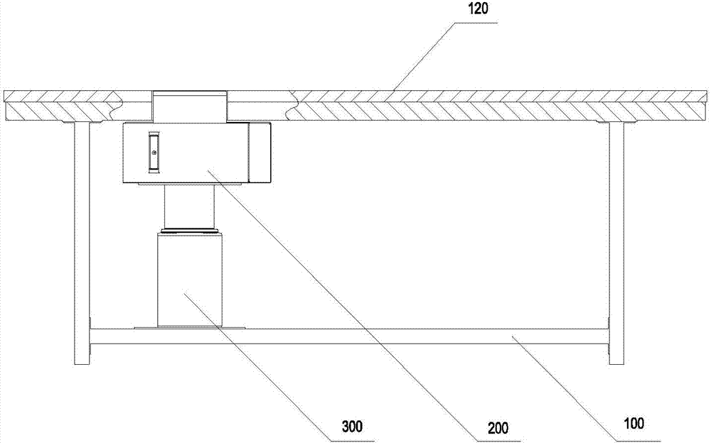
[0037] Such as figure 1 As shown, the breast ultrasound device of the present invention includes: a loading bed 100, used to support the human body, and assist the human body to perform diagnostic measurements; a scanning assembly 200, used to scan human breasts; a lifting device 300, used to scan the scanning assembly 200. Up and down to adjust the compression degree between the scanning assembly 200 and the breast.

[0038] Of course, the breast ultrasound device of the present invention may not include the lifting device 300. At this time, the degree of extrusion between the s...


PUM

PropertyMeasurementUnit
Opening angleaaaaaaaaaa

Abstract

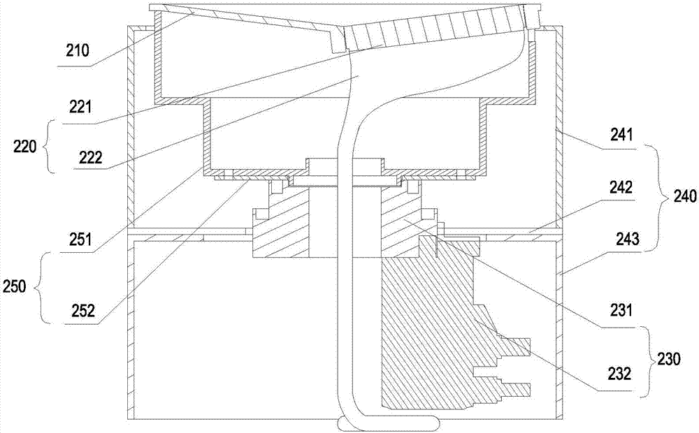
The invention relates to a breast ultrasonic device and a breast ultrasonic scanning assembly. The breast ultrasonic device comprises a scanning device and a platform which is used for supporting the scanning assembly, and is characterized in that the scanning assembly comprises a cup with a cavity, a transducer assembly and a transmission device; the transducer assembly comprises a transducer; the transducer is arranged at the bottom of the cup; the cup and the transducer assembly are connected to a power output end of the transmission device; and the transmission device drives the cup and the transducer assembly to rotate integrally. A breast can be accurately scanned and imaged full-automatically, and therefore, a doctor can be assisted to accurately judge conditions of patients and can formulate a treatment scheme in time.

Description

technical field [0001] The invention relates to a breast ultrasound device and a breast ultrasound scanning component, belonging to the technical field of ultrasound imaging. Background technique [0002] With the deterioration of the global environment and the increase of various pressures, female breast disease has become a lesion with a high incidence rate among women, and the incidence rate is increasing year by year and showing a trend of younger development. Compared with imaging diagnostic methods such as mammography, CT, and MRI, ultrasound imaging has the advantages of being able to clearly display breast mass structures and lesions, accurately identifying cystic and solid lesions, being suitable for dense breasts, and having no radiation. An inexpensive, safe, and highly accurate inspection method. At present, when examining the breast in the hospital, the general probe is mainly hand-held. In the breast examination, the probe still needs to be close to the breast...

Claims


Application Information

Patent Timeline
IPC IPC(8): A61B8/00
CPCA61B8/00A61B8/40A61B8/42A61B8/4444A61B8/4455A61B8/4461
Inventor 韩旭彭春林赵明昌王鋐
Owner CHISON MEDICAL TECH CO LTD
Learn More