Vascular tree extraction method, device and equipment and storage medium
An extraction method and vascular tree technology, applied in the field of medical image processing, can solve the problems of low efficiency, inability to provide a vascular tree extraction method, inaccurate vascular tree extraction, etc., and achieve the effect of improving effectiveness and accuracy
- Summary
- Abstract
- Description
- Claims
- Application Information
AI Technical Summary
Problems solved by technology
Method used
Image
Examples
Embodiment 1
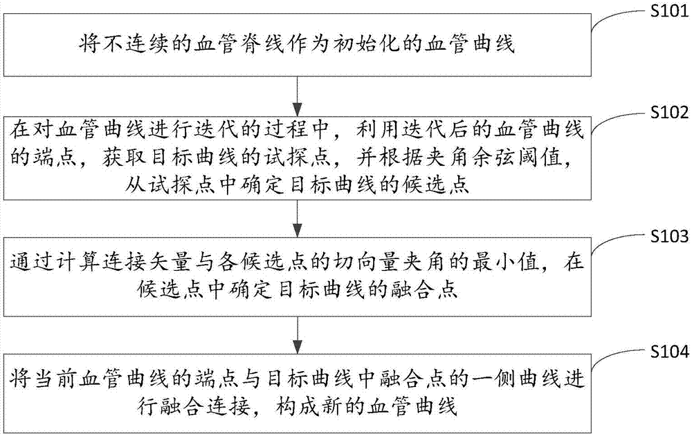
[0025] figure 1 The implementation flow of the vascular tree extraction method provided by the first embodiment of the present invention is shown. For the convenience of description, only the parts related to the embodiment of the present invention are shown, and the details are as follows:
[0026] In step S101, a discontinuous vascular ridge line is used as an initialized vascular curve.
[0027] In the embodiment of the present invention, the enhanced images of the aorta, the root point of the coronary artery, and the cardiovascular system in the CTA angiography are acquired, and the discontinuous vascular ridge line is used as the initialized vascular curve.
[0028] Further, the position coordinates and tangent vectors of each point in the blood vessel curve are obtained, and the end point attributes and curve attributes are marked on the blood vessel curve.
[0029] Specifically, the discontinuous vascular ridge line is used as the initialized vascular curve S N , S N...
Embodiment 2
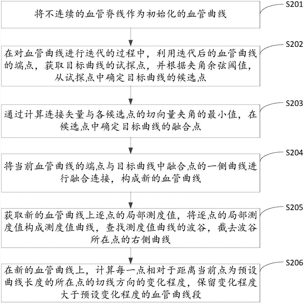
[0057] image 3 The implementation process of the vascular tree extraction method provided by the second embodiment of the present invention is shown. For the convenience of description, only the parts related to the embodiment of the present invention are shown, and the details are as follows:
[0058] In step S201, a discontinuous vascular ridge line is used as an initialized vascular curve.
[0059] In step S202, during the iterative process of the blood vessel curve, the end points of the iterated blood vessel curve are used to obtain tentative points of the target curve, and the candidate points of the target curve are determined from the tentative points according to the included angle cosine threshold.
[0060] In step S203, by calculating the minimum value of the angle between the connection vector and the tangent vector of each candidate point, the fusion point of the target curve is determined among the candidate points, and the connection vector is the connection ve...
Embodiment 3
[0074] Figure 4 It shows a schematic structural diagram of the vascular tree extraction device provided by Embodiment 3 of the present invention. For the convenience of description, only the parts related to the embodiment of the present invention are shown. The vascular tree extraction device includes:
[0075] The initialization unit 31 is configured to use discontinuous vascular ridges as initialized vascular curves.
[0076] In the embodiment of the present invention, the enhanced images of the aorta, the root point of the coronary artery, and the cardiovascular system in the CTA angiography are acquired, and the discontinuous vascular ridge line is used as the initialized vascular curve.
[0077] Further, the position coordinates and tangent vectors of each point in the blood vessel curve are obtained, and the end point attributes and curve attributes are marked on the blood vessel curve.
[0078] Specifically, the discontinuous vascular ridge line is used as the initia...
PUM
 Abstract
Description
Claims
Application Information
 - R&D
- Intellectual Property
- Life Sciences
- Materials
- Tech Scout
- Unparalleled Data Quality
- Higher Quality Content
- 60% Fewer Hallucinations
Browse by: Latest US Patents, China's latest patents, Technical Efficacy Thesaurus, Application Domain, Technology Topic, Popular Technical Reports.
© 2025 PatSnap. All rights reserved.Legal|Privacy policy|Modern Slavery Act Transparency Statement|Sitemap|About US| Contact US: help@patsnap.com


