Pulmonary nodule locating system and method for 3D pulmonary surface projection

A positioning system and pulmonary nodule technology, applied in the field of medical image processing, can solve the problems of increasing patient pain, invasive operation, difficult to remove, etc., and achieve the effect of reducing pain

Inactive Publication Date: 2017-11-24
郭明
View PDF4 Cites 12 Cited by
  • Summary
  • Abstract
  • Description
  • Claims
  • Application Information

AI Technical Summary

Problems solved by technology

At present, preoperative puncture positioning needle (Hook-wire) is used for preoperative positioning of small pulmonary nodules. The main disadvantage is that it is an invasive operation, which increases the pain of patients. If abnormalities occur after anesthesia, patients cannot tolerate it. After undergoing surgery, the positioning needle that has been punctured and fixed on the lung tissue is difficult to remove without surgery, and the patient cannot tolerate the continued surgery and is very passive

Method used


Image

  • Pulmonary nodule locating system and method for 3D pulmonary surface projection

Examples


Embodiment 1

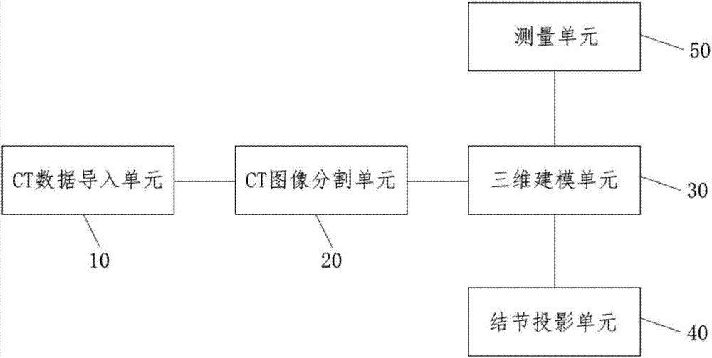
[0030] refer to figure 1 As shown, the present invention discloses a 3D lung surface projection pulmonary nodule positioning system, including a CT data import unit 10, a CT image segmentation unit 20, a three-dimensional modeling unit 30, a nodule projection unit 40 and a measurement unit 50, wherein:

[0031] The CT data import unit 10 is used for importing CT data, and the CT data includes lung CT images, patient information, hospital information, CT machine model information and CT slice accuracy information. The CT data import unit 10 has a CT data preview module and a lung CT image preprocessing module, the CT data preview module is used to view relevant content of the CT data, and the lung CT image preprocessing module is used to judge whether the lung CT image is inverted, And adjust the inverted lung CT image to the correct orientation.

[0032] The CT image segmentation unit 20 includes a lung tissue and organ segmentation module, a nodule segmentation module, a blo...

Embodiment 2

[0037] The present invention discloses a 3D lung surface projection lung nodule positioning method, which is implemented based on the 3D lung surface projection lung nodule positioning system of Embodiment 1. The method includes the following steps:

[0038] S1. Using the CT data import unit 10 to import CT data.

[0039] S2. Using the CT image segmentation unit 20 to segment the lung tissues, organs, nodules, and blood vessels on the lung CT image, and segment the lung tissues and organs according to the blood vessels. Step S2 specifically includes the following steps:

[0040] S21. Segmentation of lung tissue and organ, selecting the position of lung tissue and organ on the lung CT image through human-computer interaction, and automatically segmenting the lung tissue and organ. The position of the lung tissue and organ on the lung CT image is selected by setting the seed point through human-computer interaction, and the computer automatically segments the lung tissue and or...


PUM


Abstract

The invention discloses a pulmonary nodule locating system for 3D pulmonary surface projection. The system comprises a CT data importing unit, a CT image segmentation unit, a 3D modeling unit, a nodule projection unit and a measurement unit, wherein the CT data importing unit used for importing CT data; the CT image segmentation unit comprises a pulmonary tissue organ segmentation module, a nodule segmentation module, a blood vessel segmentation module and a segmentation module; the 3D modeling unit is used for building 3D models of a pulmonary tissue organ, a nodule and a blood vessel of each segment, and fusing the models into a pulmonary tissue 3D model; the nodule projection unit is used for building an expansion model of the pulmonary nodule, and the expansion model is the extension of the pulmonary nodule to the pulmonary surface; and the measurement unit is used for measuring a distance between the expansion model and anatomical characteristics identifiable for naked eyes on the surface of the pulmonary tissue 3D model. The invention furthermore discloses a pulmonary tissue model 3D printing method. According to the system and the method, the pulmonary nodule can be intuitively and accurately located on the premise of no traumas, so that the pains of patients are relieved and the pulmonary nodule wedge resection operation under a thoracoscope is more accurate, safer and more popular.

Description

technical field [0001] The invention relates to the technical field of medical image processing, in particular to a 3D lung surface projection pulmonary nodule positioning system and method. Background technique [0002] Lung cancer is one of the most common malignant tumors. The morbidity and mortality of lung cancer are on the rise all over the world, especially in developing countries. The incidence of lung cancer in my country is increasing by 26.9% every year, and lung cancer has become the first cause of death from malignant tumors in my country. The treatment effect of lung cancer is closely related to the stage of cancer when it is discovered. Clinical studies in thoracic surgery have shown that the 5-year survival rate of AIS and MIA treated with minimally invasive surgery such as wedge resection or segmentectomy is close to 100%; even for stage I lung cancer, the 10-year survival rate after surgical treatment can reach more than 90%. Therefore, the focus of break...

Claims


Application Information

Patent Timeline
Patent Type & Authority Applications(China)
IPC IPC(8): G06T7/11G06T19/00
CPCG06T7/11G06T19/00G06T2207/10081G06T2207/30064G06T2207/30096
Inventor 郭明杨清杰胡蒙孙晓雁包传恩解百宜黄晓媚
Owner 郭明
Why Eureka
Try Eureka