
Image processing device, living body observation device and image processing method

An image processing device and image technology, applied in image data processing, image enhancement, image analysis, etc., can solve problems such as difficult to observe, difficult to emphasize display of nerves, damage to nerves, etc., and achieve the effect of reducing risks

Active Publication Date: 2019-05-07
OLYMPUS CORP
View PDF6 Cites 0 Cited by
  • Summary
  • Abstract
  • Description
  • Claims
  • Application Information

AI Technical Summary

Problems solved by technology

[0004] However, although narrow-band light observation can emphasize blood vessels, it is difficult to emphasize nerves
[0005] For example, when nerves are preserved in total rectal resection or total prostatectomy, it is necessary to expose the target organ in order not to injure the nerves distributed so as to surround the target organ when resecting the target organ. However, thin nerves with a diameter of 50 to 300 μm are white or transparent, so it is difficult to observe even with magnified observation using a laparoscope
Therefore, doctors have to rely on experience or feeling to perform surgery, and there is a disadvantage that the possibility of nerve damage is high.

Method used


Image

  • Image processing device, living body observation device and image processing method
  • Image processing device, living body observation device and image processing method
  • Image processing device, living body observation device and image processing method

Examples


no. 1 Embodiment approach 〕

[0070] An image processing unit (image processing device), a living body observation device having the image processing unit, and an image processing method using the image processing unit and the living body observation device according to the first embodiment of the present invention will be described below with reference to the drawings.

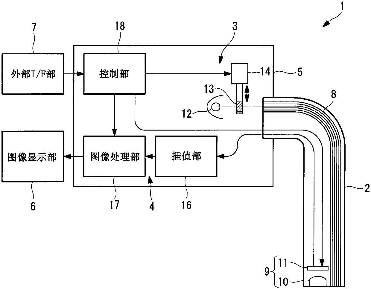
[0071] The living body observation device 1 of the present embodiment is an endoscope, such as figure 1 As shown, it has: an insertion part 2 inserted into the living body; a main body part 5, which has a light source part (illumination part) 3 and a signal processing part 4 connected to the insertion part 2; an image display part (display part) 6, It displays an image generated by the signal processing section 4; and an external interface section (hereinafter referred to as "external I / F section") 7 for input from an operator.

[0072] The insertion unit 2 has an illumination optical system 8 for irradiating a subject with light input fr...

no. 2 Embodiment approach 〕

[0171] Next, an image processing unit (image processing device), a living body observation device including the image processing unit, and an image processing method according to a second embodiment of the present invention will be described below with reference to the drawings.

[0172] In the description of the present embodiment, parts having the same configurations as those of the image processing unit 17 , the living body observation device 1 , and the image processing method of the first embodiment described above are given the same reference numerals and their descriptions are omitted.

[0173] In the first embodiment, a color CCD is used as the imaging element 11, and image signals of three channels are acquired simultaneously. The living body observation device 50 of the present embodiment replaces this case, such as Figure 13 As shown, a monochromatic CCD is used as the imaging element 51, and instead of the short-wavelength cut filter 13 and the linear motion mecha...


PUM


Abstract

Allows the surgeon to grasp areas where fat detection cannot be detected correctly due to the influence of blood or other interference, reducing the risk of nerve damage. An image processing unit is provided, comprising: a fat area setting unit (32) that detects fat area information indicating a fat area where fat exists in a biopsy image; a fat area reliability calculation unit (37) that calculates the reliability of the fat area information detected by the area setting unit (32); and the display mode setting unit which displays the fat area whose calculation reliability calculated by the fat area reliability calculation unit (37) is lower than the reference reliability as a reference. The fat region indicated by the region information is processed into a display format that can be distinguished from the surrounding region.

Description

technical field [0001] The present invention relates to an image processing device, a living body observation device and an image processing method. Background technique [0002] Conventionally, narrow-band light observation (NBI) for emphasizing and displaying capillaries on the mucosal surface and the like by irradiating illumination light of a narrowed-band wavelength that is easily absorbed by hemoglobin contained in blood is known (see, for example, Patent Document 1). [0003] This narrow-band light observation is expected as an alternative observation method for pigment dispersion widely performed for detailed diagnosis of the esophagus region or observation of pockmarked images (glandular structure) of the large intestine. Based on the reduction of examination time and unnecessary biopsies, It can be expected to contribute to the improvement of inspection efficiency. [0004] However, narrow-band light observation is capable of emphasizing blood vessels, but it is d...

Claims


Application Information

Patent Timeline
Patent Type & Authority Patents(China)
IPC IPC(8): A61B1/04
CPCA61B1/00045A61B1/0638A61B1/000094A61B1/044A61B5/4872A61B5/7203A61B5/7221A61B5/7278A61B2576/00G06T7/0012G06T2207/10068G06T2207/30101
Inventor 森田惠仁
Owner OLYMPUS CORP
Learn More