Method and device for detection of gene fusion

A technology of gene fusion and detection method, applied in special data processing applications, instruments, electrical digital data processing, etc., to achieve the effect of low false positives and high accuracy

Active Publication Date: 2017-12-15
武汉安至康永医疗科技有限公司
View PDF4 Cites 7 Cited by
  • Summary
  • Abstract
  • Description
  • Claims
  • Application Information

AI Technical Summary

Problems solved by technology

However, in the detection of tumor DNA in blood, due to the extreme concentration (<1%), most of the DNA is normal DNA, which cannot be identified by conventional assembly methods

Method used


Image

  • Method and device for detection of gene fusion

Examples


Embodiment approach

[0023] According to a typical embodiment of the present invention, the detection method includes S0: repeating sequence filtering, and the repeating sequence filtering step includes: S01, designing a simulated base sequence of 30-75 bp, aligning with the reference genome sequence, finding It is found that the coverage of the simulated base sequence on the reference gene is significantly higher than that of the surrounding sequence, and the region is marked as a highly repetitive region; S02, before performing sequence comparison in step S1 and / or step S42, first The highly repetitive regions in the reference genome are filtered to improve the accuracy of the sequence alignment. Therefore, when the gene sequence to be detected is compared with the reference genome, repetitive sequences can be avoided, so as to improve the efficiency and accuracy of the alignment.

[0024] According to a typical embodiment of the present invention, S41 uses the K-mer algorithm to concatenate to ...

Embodiment 1

[0033] An embodiment of the method for detecting gene fusion of the present invention, wherein:

[0034] Detection object: tumor circulating DNA.

[0035] The concentration of tumor DNA in blood is very low, and most of the sequencing sequences that can be detected by DNA sequencing extracted from blood are derived from tissue DNA sequences. Therefore, in this embodiment, tumor DNA is first identified from tissue DNA, and then gene fusion is identified.

[0036] Detection method: including the following steps:

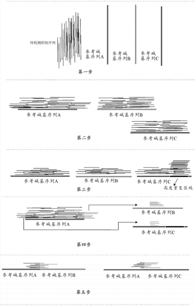
[0037] like figure 1 As shown, the first step: the short lines clustered together on the left are the short sequences to be sequenced; the three lines on the right are the reference sequences of the three genes (named A, B, C, respectively);

[0038] Step 2: Use sequence alignment software to align the short sequence to be sequenced to the reference gene sequence;

[0039] Step 3: Design a 30-75bp simulated base sequence, and use the 30-75bp simulated base sequence...

Embodiment 2

[0048] Three standard products with gene fusion were ordered from the invitrogen website, and after being processed by the gene fusion detection device of the present invention, all the gene fusion sequences were identified, and the positions of the gene fusions were consistent with the results of the standard products; the gene fusion of the standard products The position of the break is fixed (the standard does not give the fusion position), not random, so as long as the detected fusion gene sequence is correct, the position of the gene fusion is also consistent with the standard. The detected frequencies of gene fusions (Table 1 below) were also approximately the same.

[0049] Table 1 Detection effect of the gene fusion detection device of the present invention

[0050] sample name

gene fusion

Standard fusion frequency

Detected fusion frequency

HD664

ALK-EML4

50.00%

43.58%

HD753

ROS1-SLC32A

5.00%

4.20%

HD753...


PUM


Abstract

The invention discloses a method for detection of gene fusion. The method includes the following steps that 1, the sequences of to-be-detected gene fragments are provided, and sequence comparison with a reference genome is conducted; 2, the to-be-detected gene fragments which are partially same as the sequence of the reference genome are extracted, and consensus sequence portions are labeled; 3, sequence comparison is conducted again on the to-be-detected gene fragments extracted in the step 2 and the reference genome, and if the obtained consensus sequence is not same as the consensus sequence labeled in the step 2, it is speculated that the to-be-detected gene fragments extracted in the step 2 have a gene fusion phenomenon; 4, the authenticity of gene fusion is verified. At the same time, the method and the device further discloses a device for the detection of gene fusion. By means of the method and the device, the gene fusion sequence (less than 1%) with the extremely low concentration can be accurately detected out, and the method and the device are more sensitive to the gene fusion sequence high in fusion concentration.

Description

technical field [0001] The invention relates to the technical field of gene detection, in particular to a gene fusion detection method and device. Background technique [0002] Cancer cells release DNA from their own cells into the blood when they die. Because different cancer cells are different, the DNA released into the blood contains DNA mutations that are unique to different cancers. The DNA of these tumor cells released into the blood is called tumor circulating DNA (cell tumor DNA, ctDNA). But the amount of these DNAs in blood is very low, generally less than 1%. Previous approaches to large-scale variant detection typically employed high-coverage next-generation sequencing (<500-fold) to sequence tissue DNA, followed by PCR validation. The frequencies of DNA variants detected by these techniques are generally greater than 1%. These have changed with the advent of digital PCR, which can detect variants with frequencies greater than 0.01%, so these tiny amounts o...

Claims


Application Information

Patent Timeline
Patent Type & Authority Applications(China)
IPC IPC(8): G06F19/22
CPCG16B30/00
Inventor 龚浩车健为
Owner 武汉安至康永医疗科技有限公司
Why Eureka
Try Eureka