A tissue classification method and device based on cardiovascular ivoct images

A classification method and cardiovascular technology, applied in image analysis, image enhancement, image data processing, etc., can solve problems such as inability to truly restore tissue structure, image information loss, and time-consuming, so as to achieve good image display effect and compensate for image information loss , High reduction effect

Active Publication Date: 2021-11-19
中科微光医疗研究中心(西安)有限公司
View PDF3 Cites 0 Cited by
  • Summary
  • Abstract
  • Description
  • Claims
  • Application Information

AI Technical Summary

Problems solved by technology

[0003] Normal arteries have a uniform layered structure consisting of intima, media and adventitia, but when blood vessels are diseased, different types of tissues will be contained in the blood vessels. Therefore, it is necessary to classify and detect these different tissues. However, , until now, the detection and classification of these tissues mainly rely on manual work, which is very time-consuming
[0004] A convolutional network for biomedical image segmentation is proposed in the prior art. This method uses a contraction path to capture content, an enlargement path to precisely locate, and two paths to form a U-shape, called U-Net. The final output image resolution of U-Net will be slightly smaller than the original image, resulting in the loss of image information and unable to truly restore the organizational structure

Method used


Image

  • A tissue classification method and device based on cardiovascular ivoct images
  • A tissue classification method and device based on cardiovascular ivoct images
  • A tissue classification method and device based on cardiovascular ivoct images

Examples


Embodiment 1

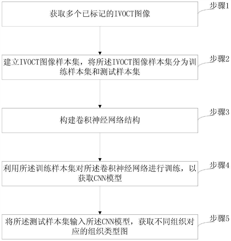
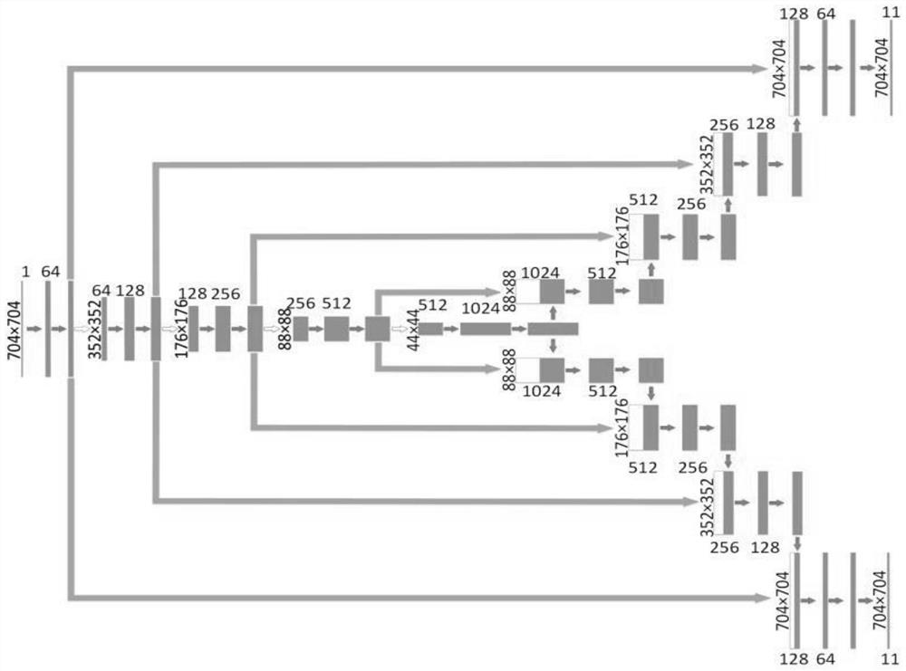
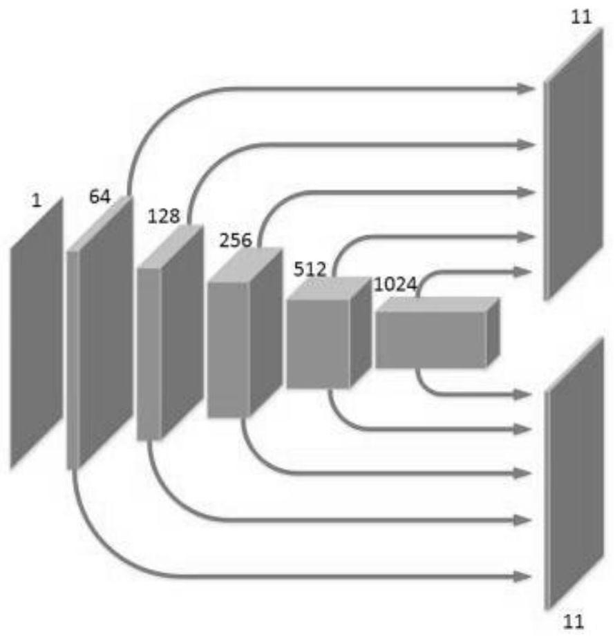
[0043] Such as Figure 1-Figure 5 as shown, figure 1 A flow chart of the tissue classification method provided by the embodiment of the present invention; figure 2 The structural diagram of the convolutional neural network provided by the embodiment of the present invention; Fig. 3 (a) is a schematic diagram of the CNN model provided by the embodiment of the present invention; Fig. 3 (b) is a virtual diagram of the use of the CNN model provided by the embodiment of the present invention ; Fig. 4 (a) is the cardiovascular IVOCT image of the input end input of the CNN model provided by the embodiment of the present invention; Fig. 4 (b) is the tissue segmentation diagram of the first output end output of the CNN model provided by the embodiment of the present invention; Fig. 4 (c) is the organizational boundary map that the second output terminal output of the CNN model provided by the embodiment of the present invention; Figure 5 The combined cardiovascular tissue classific...


PUM


Abstract

The present invention relates to a method and device for tissue classification based on cardiovascular IVOCT images, wherein the method includes: step 1, acquiring a plurality of marked IVOCT images; step 2, establishing an IVOCT image sample set, and storing the IVOCT images The sample set is divided into a training sample set and a test sample set; Step 3, constructing a convolutional neural network structure; Step 4, using the training sample set to train the convolutional neural network to obtain a CNN model; Step 5, incorporating The test sample set is input into the CNN model to obtain tissue type maps corresponding to different organizations. In the embodiment of the present invention, the CNN model has two output terminals, which respectively display the outline and internal structure of the tissue, which solves the problem that in the prior art, the resolution of the output image is slightly small, and some image information is lost, resulting in tissue boundaries. Shows unclear technical issues.

Description

technical field [0001] The invention belongs to biological tissue imaging technology, in particular to a method and device for tissue classification based on cardiovascular IVOCT images. Background technique [0002] A biopsy is a common medical test in which a pathologist looks at a sample of tissue taken from a subject under a microscope to determine the nature or extent of disease. Typically, tissue needs to be cut into very thin sections and stained before it can be viewed under a microscope. Optical coherence tomography (OCT) is an alternative to non-destructive optical imaging modalities that can provide high-resolution 3D images of tissue from biopsy samples without staining. Optical coherence microscopy (OCM) combines the advantages of OCT and confocal microscopy to provide high-resolution cellular images. [0003] Normal arteries have a uniform layered structure composed of intima, media and adventitia, but when blood vessels are diseased, different types of tissu...

Claims


Application Information

Patent Timeline
Patent Type & Authority Patents(China)
IPC IPC(8): G06T7/00G06T7/12G06K9/62G06N3/04G06N3/08
CPCG06T7/0012G06T7/12G06T2207/30101G06T2207/20081G06T2207/20084G06T2207/10101G06T2207/10061G06F18/24G06F18/214
Inventor 朱锐曹一挥薛婷
Owner 中科微光医疗研究中心(西安)有限公司
Why Eureka
Try Eureka