Auxiliary diagnostic system for interpreting medical image features based on deep learning method

A deep learning and medical image technology, applied in neural learning methods, image enhancement, image analysis, etc., can solve problems such as poor quality of ultrasound images, accuracy of auxiliary diagnosis, and impact of automation

Inactive Publication Date: 2018-07-06
ZHEJIANG DE IMAGE SOLUTIONS CO LTD
View PDF7 Cites 68 Cited by
  • Summary
  • Abstract
  • Description
  • Claims
  • Application Information

AI Technical Summary

Problems solved by technology

However, the inherent imaging mechanism makes the quality of clinically acquired ultrasound images poor, which affects the accuracy and automation of auxiliary diagnosis. Therefore, the current most lesions in segmented ultrasound images are semi-automatic segmentation based on active contours, and the classification is mainly It is to manually select features

Method used


Image

  • Auxiliary diagnostic system for interpreting medical image features based on deep learning method
  • Auxiliary diagnostic system for interpreting medical image features based on deep learning method
  • Auxiliary diagnostic system for interpreting medical image features based on deep learning method

Examples


Embodiment Construction

[0079] Below in conjunction with accompanying drawing and specific embodiment the present invention is described in further detail:

[0080] The following examples can enable those skilled in the art to understand the present invention more comprehensively, but do not limit the present invention in any way.

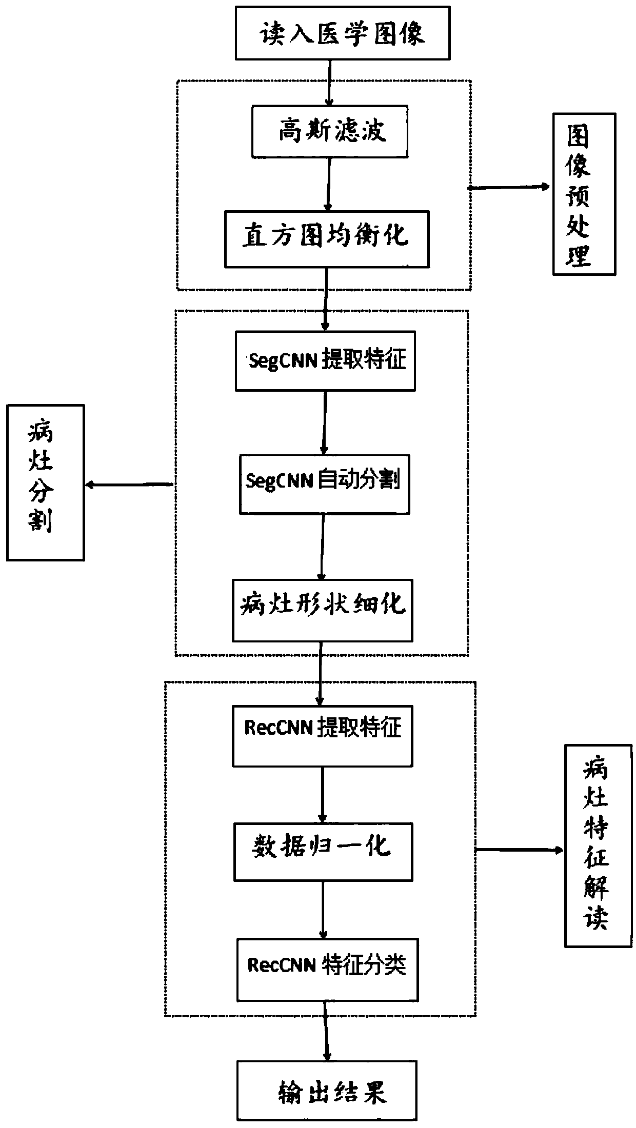
[0081] Such as figure 1 As shown, an auxiliary diagnosis system for interpreting medical image features based on deep learning method, including the following steps:

[0082] 1. Read the medical imaging data of the lesion:

[0083] Read medical images of lesions, including at least 10,000 images of benign lesions and at least 10,000 images of malignant lesions; images can be in image format or standard dicom images.

[0084] 2. Preprocessing of medical images:

[0085] The image of the lesion read in the first process is grayed first, and the gray value of the surrounding pixels is used to remove the mark made by the doctor for measuring the nodule related quantity in ...


PUM


Abstract

The invention relates to the field of auxiliary medical diagnosis and aims to provide an auxiliary diagnosis system for interpreting medical image features based on the deep learning method. The system comprises the following steps of: reading medical image data of a lesion and preprocessing the medical image data; selecting an image, establishing a convolutional neural network architecture, automatically learning and segmenting a lesion area, and refining the shape of the lesion; constructing a CNN model of a convolutional neural network architecture to automatically interpret the characteristics of benign and malignant lesions, and acquiring an auxiliary diagnosis system for interpreting medical image features based on the depth learning method after training. The invention not only canautomatically divide the focus area by means of the depth convolution neural network, make up the deficiency that the weak boundary problem cannot be solved based on the active contour and the like, but also can automatically learn the characteristic combination extracted with the value, thereby avoiding the complex of manually selecting the feature.

Description

technical field [0001] The invention relates to the field of auxiliary medical diagnosis, in particular to an auxiliary diagnosis system for interpreting medical image features based on a deep learning method. Background technique [0002] In recent years, with the rapid development of computer technology and digital image processing technology, digital image processing technology has been more and more used in the field of auxiliary medical diagnosis. Accurate, recognition and other image processing technologies to obtain valuable medical diagnosis information, the main purpose is to make the doctor observe the lesion more directly and clearly, and provide auxiliary reference for the doctor's clinical diagnosis, which has very important practical significance. [0003] Based on medical imaging, early detection of lesions is of great significance for the identification of benign and malignant lesions, clinical treatment and surgical selection. Ultrasound examination based o...

Claims


Application Information

Patent Timeline
IPC IPC(8): G06T7/11G06K9/46G06K9/62G06T7/00G06T7/136G06N3/08
CPCG06N3/08G06T7/0012G06T7/11G06T7/136G06T2207/10132G06V10/44G06F18/2411
Inventor 胡海蓉
Owner ZHEJIANG DE IMAGE SOLUTIONS CO LTD
Why Eureka
Try Eureka