
Bone joint CT image segmentation method and system

A CT image, bone and joint technology, applied in the field of image processing, can solve the problems of time-consuming, labor-intensive, low-efficiency, etc., and achieve the effect of improving accuracy and accuracy

Inactive Publication Date: 2018-07-10
BOCO INTER TELECOM
View PDF4 Cites 11 Cited by
  • Summary
  • Abstract
  • Description
  • Claims
  • Application Information

AI Technical Summary

Problems solved by technology

[0002] At present, most of the three-dimensional segmentation methods for bone and joint CT images at home and abroad require manual positioning or manual segmentation in each CT sequence image, which is time-consuming, laborious, and inefficient.

Method used


Image

  • Bone joint CT image segmentation method and system
  • Bone joint CT image segmentation method and system
  • Bone joint CT image segmentation method and system

Examples


Embodiment Construction

[0051] The following will clearly and completely describe the technical solutions in the embodiments of the present invention with reference to the accompanying drawings in the embodiments of the present invention. Obviously, the described embodiments are only some, not all, embodiments of the present invention. Based on the embodiments of the present invention, all other embodiments obtained by persons of ordinary skill in the art without making creative efforts belong to the protection scope of the present invention.

[0052] CT scans a layer of a certain thickness of a certain part of the human body with an X-ray beam. The X-rays that pass through this layer are received by the detector and converted into visible light, which is converted from photoelectricity into an electrical signal, and then passed through an analog / digital converter ( analog / digital converter) to digital signal, input to computer for processing. The processing of image formation is like dividing the se...


PUM


Abstract

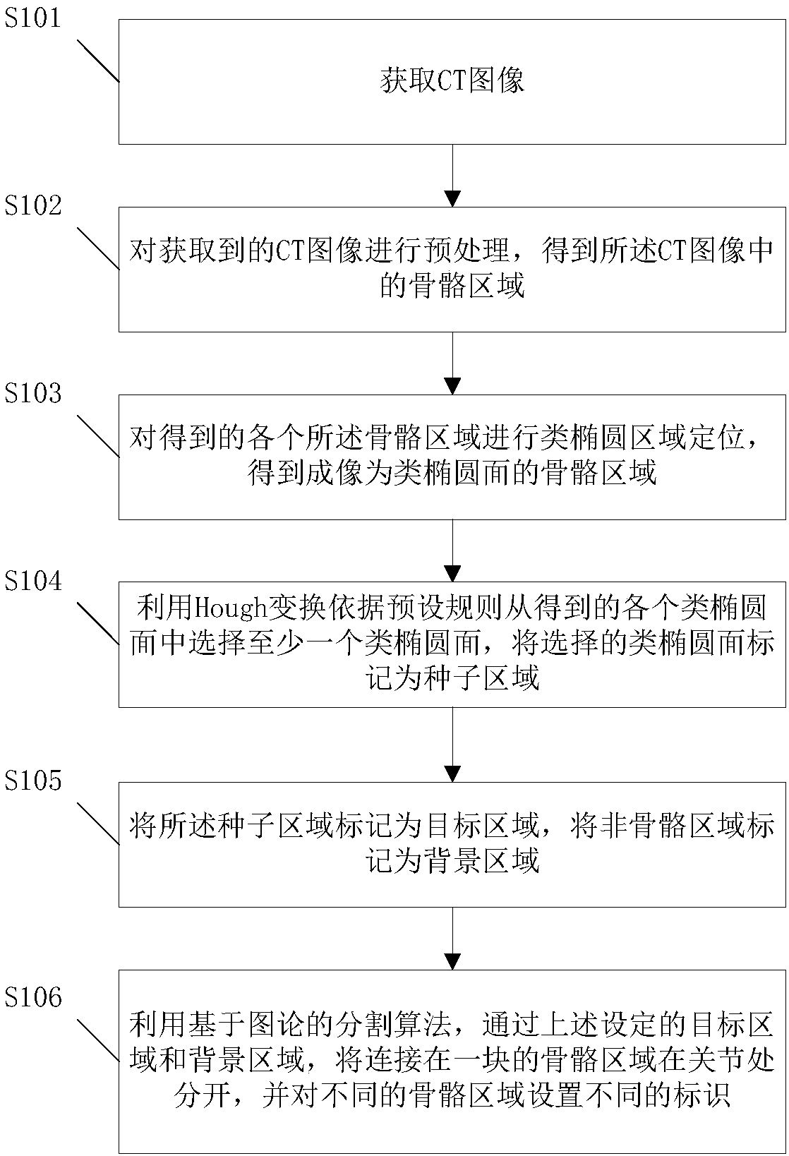
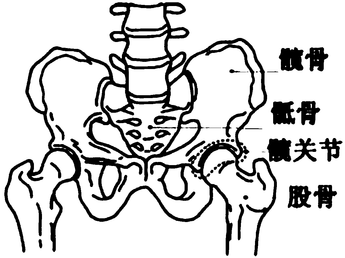
The invention provides a bone joint CT image segmentation method and system. The acquired CT image is preprocessed so as to obtain the bone areas of the CT image; quasi-elliptic area locating is performed on each obtained bone area so as to obtain the bone areas imaged as the quasi-ellipsoids; at least one quasi-ellipsoid is selected from all the obtained quasi-ellipsoids according to the preset rules, and the selected quasi-ellipsoid is marked as the seed area; and the seed area is marked as the target area, and the non-bone areas are marked as the background areas. The connected bone area isseparated at the joints through the preset target area and the background areas by using the energy minimum segmentation algorithm based on the graph theory, and different identifiers are set for different bone areas so that CT image automatic segmentation can be realized and the efficiency of CT image segmentation can be enhanced.

Description

technical field [0001] The invention relates to the technical field of image processing, in particular to a bone joint CT image segmentation method and system. Background technique [0002] At present, most of the three-dimensional segmentation methods for bone and joint CT images at home and abroad require manual positioning or manual segmentation in each CT sequence image, which is time-consuming, laborious, and inefficient. Therefore, there is an urgent need for a segmentation method for fast segmentation of joint images. Contents of the invention [0003] In view of this, an embodiment of the present invention provides a bone joint CT image segmentation method and system, so as to realize the automatic segmentation of the bone joint CT image. [0004] In order to achieve the above purpose, embodiments of the present invention provide the following technical solutions: [0005] A bone joint CT image segmentation method, comprising: [0006] performing preprocessing o...

Claims


Application Information

Patent Timeline
IPC IPC(8): G06T7/11G06T7/136
CPCG06T2207/10081G06T2207/20081G06T2207/30008
Inventor 王瑜张晓璐高翔温涛
Owner BOCO INTER TELECOM
Learn More