Electrocardiogram phonocardiogram analysis method

An analysis method and electrocardiogram technology, applied in medical science, stethoscope, sensor, etc., can solve the problems of underutilization of clinical application value of heart sounds, self-monitoring of unfavorable heart patients, and analysis of heart sounds and electrocardiogram cardiac function.

Inactive Publication Date: 2018-07-27
河南善仁医疗科技有限公司
View PDF15 Cites 8 Cited by
  • Summary
  • Abstract
  • Description
  • Claims
  • Application Information

AI Technical Summary

Problems solved by technology

[0004] There have been many attempts at home and abroad on the joint monitoring of heart sounds and ECG, but the previous cardiac function detectors were limited to the traditional digitization of cardiac auscultation, and did not fully utilize the clinical application value of heart sounds, nor did they integrate heart sounds Correlation with ECG by physiological relationship for analysis of heart function
[0005] Moreover, the existing electrocardiogram and phonocardiogram still remain at the stage of manual reading, and the information of the electrocardiogram or phonocardiogram must be analyzed only by relying on the experience of doctors. Interpretation of the electrocardiogram to determine whether there is a heart disease or a hidden danger of the disease; it is not conducive to the self-monitoring of heart disease patients at home
[0006] The portable electrocardiograph that appears now can only monitor the ECG, but cannot monitor the heart sound, and the electrocardiometer only outputs the electrocardiogram, but does not send out whether the heart is abnormal. It still depends on professionals to interpret the electrocardiogram.

Method used


Image

  • Electrocardiogram phonocardiogram analysis method
  • Electrocardiogram phonocardiogram analysis method
  • Electrocardiogram phonocardiogram analysis method

Examples


Embodiment Construction

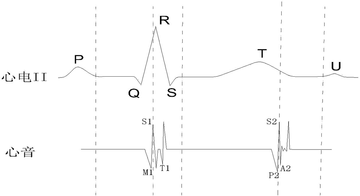
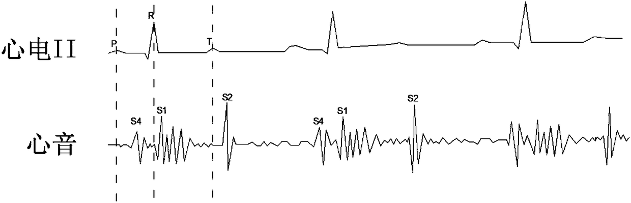
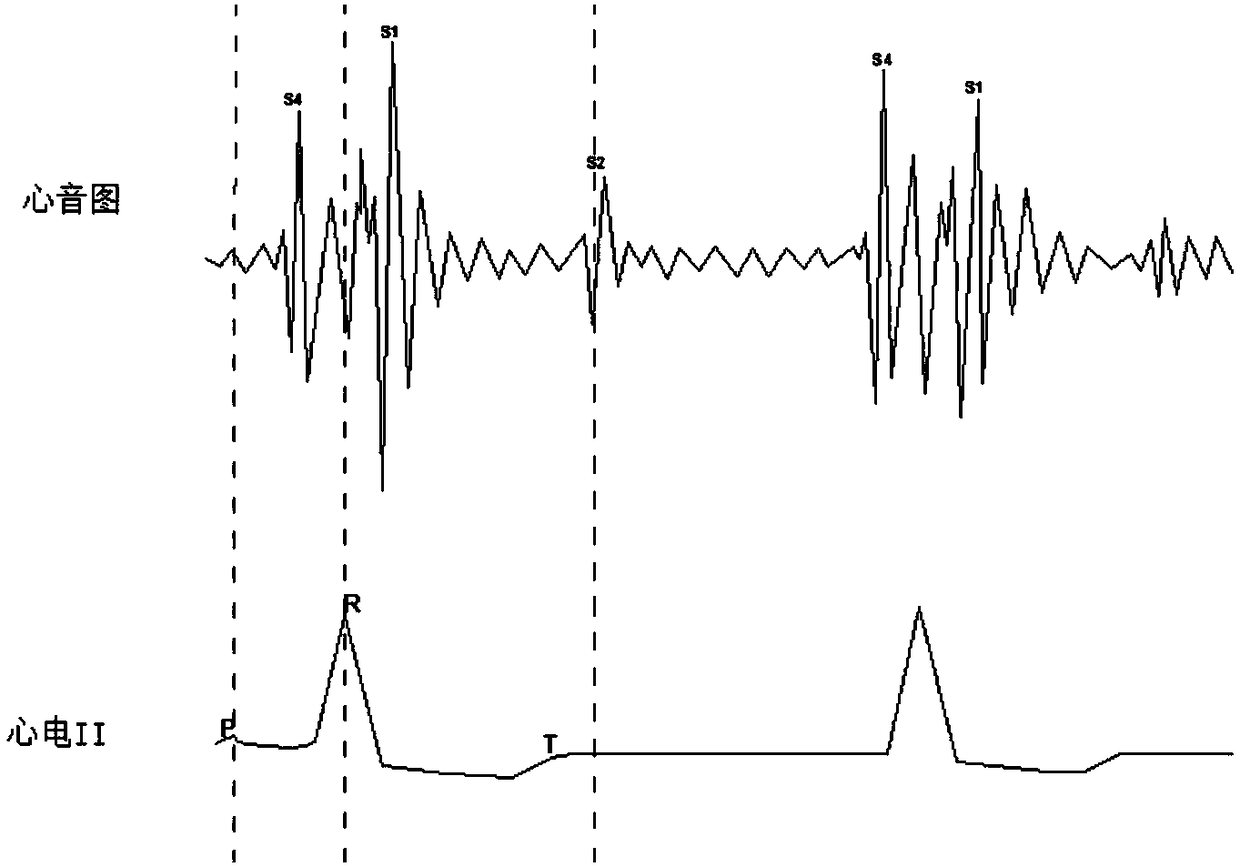
[0100] The principle of the present invention is: the electrical signal sent by the sinoatrial node is transmitted to the right atrium and the left atrium, and the excitement of the atrium is shown as the P wave of the electrocardiogram. Right ventricle, ventricular excitation is manifested as QRS complex in ECG, and ventricular repolarization is manifested as T wave in ECG. Ventricular repolarization awaits the next activation of the sinoatrial node. In the same cardiac cycle, the ECG should have P wave, QRS complex and T wave.

[0101] The generation of S1 is related to the closure of mitral valve (T1) and tricuspid valve (M1), and the generation of S2 is caused by the closure of aortic valve (A2) and pulmonary valve (P2). During one cardiac cycle, the phonocardiogram should have S1 and S2. When the cardiac cycle of the electrocardiogram does not correspond to the cardiac cycle of the phonocardiogram, it is likely that the state of the heart is abnormal.

[0102] Accordin...


PUM


Abstract

The invention provides an electrocardiogram phonocardiogram analysis method. The method comprises following steps: synchronously acquiring and synchronously recording electrocardiogram and phonocardiogram, and using the same timeline for the electrocardiogram and phonocardiogram; determining whether the P wave, R wave and T wave in the electrocardiogram can be identified; if the P wave, the R waveand the T wave in the electrocardiogram can be identified, the phonocardiogram corresponding to the RT interval is intercepted, and as the first waveform to be filtered, the phonocardiogram corresponding to the TP interval is intercepted as the second waveform to be filtered; if there is no pulse waveform in the first wave to be filtered, it is considered that S1 is missing and the heart is abnormal; if there is no pulse waveform in the second wave to be filtered, it is considered that S2 is missing and the heart is abnormal; if the electrocardio feature can not recognized in the electrocardiogram, the S1 waveform and the S2 waveform are identified from the phonocardiogram. If so, each S1 waveform and each S2 waveform are extracted; otherwise, abnormal heart condition signals are output.The invention has the advantages of being able to initially identify the function of the heart by using the matching relationship between the electrocardiographic characteristics and the heart sound characteristics, and is suitable for the advantages of home cardiac monitoring.

Description

technical field [0001] The invention relates to an analysis method of an electrocardiogram and a phonocardiogram. Background technique [0002] In the human heart, the sinoatrial node automatically and rhythmically generates current, and the current is transmitted to various parts of the heart in the order of conduction tissue, thereby causing the contraction and relaxation of cardiomyocytes. The excitement from the sinoatrial node is transmitted to the atria and ventricles in a certain way and process, causing the excitement of the whole heart. Therefore, the direction, route, sequence and time of the electrical changes occurring during the excitation of each part of the heart are regular. This bioelectrical change is reflected to the body surface through the conduction resistance value and body fluid around the heart, so that all parts of the body undergo regular electrical changes in each cardiac cycle. The electrical change curve of the heart recorded by placing the gu...

Claims


Application Information

Patent Timeline
Patent Type & Authority Applications(China)
IPC IPC(8): A61B5/0402A61B5/0472A61B7/04A61B5/366
CPCA61B5/7267A61B7/04A61B5/316A61B5/318A61B5/366
Inventor 边俊杰邹文初邹旭辉
Owner 河南善仁医疗科技有限公司
Why Eureka
Try Eureka