Method for generating positioning model and method for processing sagittal image of spine

A sagittal and spine technology, applied in the field of image processing, can solve problems such as a large amount of labor, error-prone, and poor image quality, and achieve the effects of assisting disease diagnosis, avoiding errors, and saving manpower and time costs

Active Publication Date: 2020-09-29
LONGWOOD VALLEY MEDICAL TECH CO LTD
View PDF5 Cites 0 Cited by
  • Summary
  • Abstract
  • Description
  • Claims
  • Application Information

AI Technical Summary

Problems solved by technology

However, there are still some shortcomings in the process of using MRI images to diagnose spinal diseases. For example, under the condition of interference, the image quality is not good, and the positioning of bone marker points is still based on the naked eye judgment, which needs to rely on the experience of doctors. and image quality, this approach is both error-prone and labor-intensive

Method used


Image

  • Method for generating positioning model and method for processing sagittal image of spine
  • Method for generating positioning model and method for processing sagittal image of spine
  • Method for generating positioning model and method for processing sagittal image of spine

Examples


Embodiment approach

[0089] According to one embodiment, implementing the first prediction model 900 and the second prediction model 1000 alone can obtain a prediction result about whether the input image of the intervertebral disc area is normal, that is, the prediction of whether the intervertebral disc included in the image of the intervertebral disc area is healthy result. In some embodiments according to the present invention, when the first probability is not less than 0.3, predict the health of the intervertebral disc included in the input intervertebral disc region image; when the second probability is not less than 0.5, predict the input intervertebral disc region image contains intervertebral disc health. The above prediction results are used as a reference to assist professional doctors to complete the diagnosis of the sagittal image of the spine.

[0090] In another embodiment of the present invention, the prediction results of the first prediction model 900 and the second prediction ...


PUM


Abstract

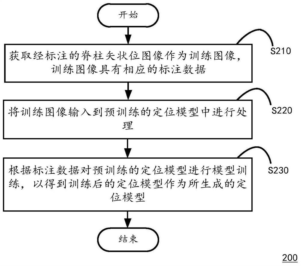
The invention discloses a method for generating a positioning model, a method for performing bone positioning on a spine sagittal image, and a computing device for executing the above method. Wherein, the method for generating the localization model includes the steps of: obtaining a marked spine sagittal image as a training image, and the training image has corresponding labeled data; inputting the training image into a pre-trained localization model for processing, and the localization model includes volume A product processing layer, a classification processing layer and a regression processing layer, wherein the convolution processing layer performs convolution, activation and pooling processing on the input image to output at least one bone positioned, and the classification processing layer and the regression processing layer pair The located bones are subjected to classification and regression processing to output the predicted probability that the bone belongs to the sacrum and the predicted position of the bone; model training is performed on the pre-trained positioning model according to the labeled data to obtain the trained positioning model.

Description

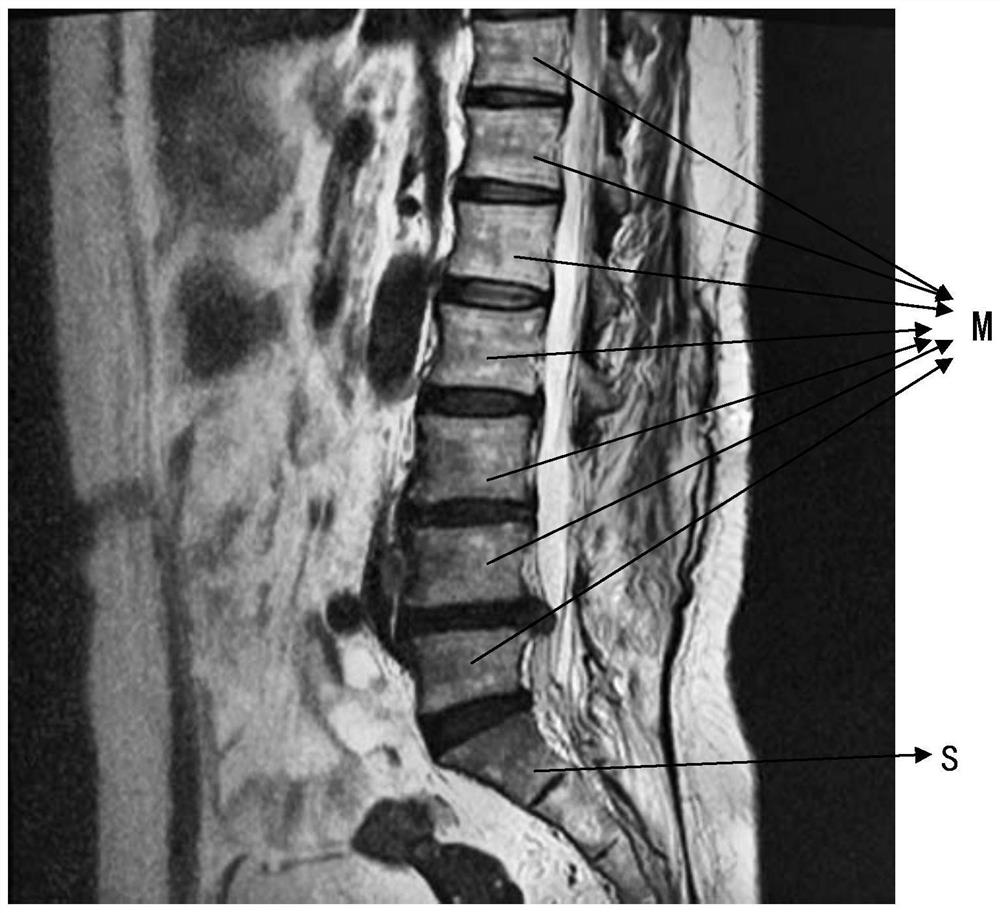
technical field [0001] The invention relates to the technical field of image processing, in particular to a method for generating a positioning model and a method for processing a spine sagittal image. Background technique [0002] With the rapid development of computer technology and image processing technology, the use of computer technology to assist orthopedic precision surgery is gradually increasing. The most common application is Magnetic Resonance Imaging (MRI) imaging, from which a variety of physical property parameters of matter can be obtained, such as proton density, spin-lattice relaxation time T1, spin-spin Spin relaxation time T2, diffusion coefficient, magnetic susceptibility coefficient, chemical shift, etc., in order to apply it to the imaging diagnosis of various systems of the whole body. One of the main applications is spinal imaging to obtain sagittal images of the human spine, which can then be used in the diagnosis of various spinal diseases, such a...

Claims


Application Information

Patent Timeline
Patent Type & Authority Patents(China)
IPC IPC(8): G06T7/00G06T7/73A61B5/055
CPCA61B5/055A61B5/4566G06T7/0012G06T7/73A61B5/7267G06T2207/30012G06N3/045
Inventor 张逸凌刘星宇安奕成张云东
Owner LONGWOOD VALLEY MEDICAL TECH CO LTD
Why Eureka
Try Eureka