A single-molecule positioning microscopic imaging method, optical component and imaging system

A technology for positioning microscopy and imaging methods, which is applied in the direction of material analysis, scientific instruments, and analytical materials through optical means. It can solve the problems of lower resolution and inability to maintain high axial resolution, and achieve large depth of field detection. Difficulties, the effect of improving the axial positioning range and resolution

Active Publication Date: 2021-02-19
SHENZHEN UNIV
View PDF6 Cites 0 Cited by
  • Summary
  • Abstract
  • Description
  • Claims
  • Application Information

AI Technical Summary

Problems solved by technology

However, in the process of 3D scanning, different levels of information will affect each other, resulting in background noise and fluorescence bleaching, reducing resolution
Some researchers propose to use deformable gratings to detect multi-layered samples, but since the energy of deformed gratings is mainly distributed in the 0th order and ±1st order of diffraction, this method can only simultaneously image nine different layers in the cell at most. Therefore, Inability to maintain high axial resolution while achieving large axial detection range

Method used


Image

  • A single-molecule positioning microscopic imaging method, optical component and imaging system
  • A single-molecule positioning microscopic imaging method, optical component and imaging system
  • A single-molecule positioning microscopic imaging method, optical component and imaging system

Examples


Embodiment Construction

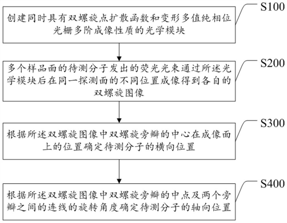
[0062] The purpose of the present invention is to provide a single-molecule positioning microscopic imaging method, optical components and imaging system, which can image molecular information at multiple levels in the sample in the form of a double helix at different positions on the same detection surface, without scanning In the case of improving the axial positioning range and resolution of double helix point spread function engineering, it solves the problem of large depth of field detection in single molecule positioning and tracking technology in living cells.

[0063] In order to make the object, technical solution and effect of the present invention more clear and definite, the present invention will be further described in detail below with reference to the accompanying drawings and examples. It should be understood that the specific embodiments described here are only used to explain the present invention, not to limit the present invention.

[0064] see figure 1 ,...


PUM


Abstract

The invention discloses a single-molecule positioning micro-imaging method, an optical assembly and an imaging system. The method comprises the following steps: establishing an optical module with a double helix point dispersion function and a deformed multi-value pure phase grating multi-order imaging property; after fluorescence beams emitted by to-be-tested molecules of multiple sample surfacesare emitted through the optical module, carrying out imaging at different positions of the same detection surface, so as to respectively generate double helix images; determining transverse positionsof the to-be-tested molecules according to positions of the centers of double helix sidelobes in the double helix images on the imaging surfaces; and determining the axial positions of the to-be-tested molecules according to the middle points of the double helix sidelobes in the double helix images and a rotation angle of a connection line between the two sidelobes. The molecular information of multiple layers in the sample can be imaged at different positions of the same detection surface in a double helix manner, so that the axial positioning range and resolution ratio of a double helix point spread function can be improved without scanning, and the problem of large field depth detection in single molecule positioning and tracing techniques in living cells is solved.

Description

technical field [0001] The invention relates to the technical field of super-resolution microscopic imaging, in particular to a single-molecule positioning microscopic imaging method, an optical component and an imaging system. Background technique [0002] Today, with the rapid development of life science and technology, in order to further understand and study the interaction between living organisms and the mechanism of disease, people urgently need to obtain more accurate structural information inside cells. However, due to the existence of the optical diffraction limit, the resolution of conventional optical microscopes can only reach about 200nm, which is difficult to meet the needs of modern biomedicine. In recent years, single-molecule localization super-resolution fluorescence microscopy has emerged, such as photosensitive localization microscopy (PLM), stochastic optical reconstruction microscopy (STORM), fluorescence photosensitive localization microscopy (FPLM), ...

Claims


Application Information

Patent Timeline
Patent Type & Authority Patents(China)
IPC IPC(8): G01N21/64G01N21/01
CPCG01N21/01G01N21/6402G01N21/6486
Inventor 于斌王美昌李四维曹慧群林丹樱屈军乐
Owner SHENZHEN UNIV
Why Eureka
Try Eureka