Method for predicting growth of coronary atherosclerotic plaque

A technology for coronary atherosclerosis and prediction methods, applied in the field of medical imaging, can solve the problems of limited penetration depth, inability to build solid models, inability to penetrate blood vessels, etc., to optimize prediction models and prevent sudden cardiovascular disease. Effect

Inactive Publication Date: 2018-12-28
SOUTHEAST UNIV
View PDF8 Cites 22 Cited by
  • Summary
  • Abstract
  • Description
  • Claims
  • Application Information

AI Technical Summary

Problems solved by technology

Although the OCT image has high resolution, its penetration depth is limited, only about 1-2 mm, so it cannot penetrate the entire blood vessel, and can only see the structure near the blood vessel lumen, so it can only establish a fluid model, and cannot establish a complete solid Model
In view of these characteristics of the OCT image, although it can

Method used


Image

  • Method for predicting growth of coronary atherosclerotic plaque
  • Method for predicting growth of coronary atherosclerotic plaque
  • Method for predicting growth of coronary atherosclerotic plaque

Examples


Embodiment 1

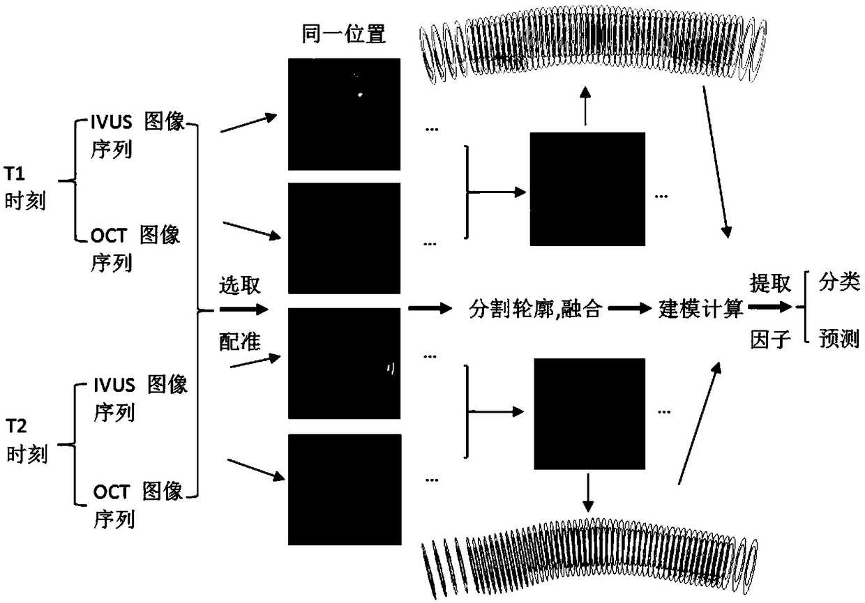
[0051] Next, we use the IVUS and OCT data at T1 and T2 to describe the method for predicting the growth of Amin's coronary atherosclerotic plaque.

[0052] Step 1: Obtain the IVUS and OCT data of the same patient's baseline (baseline, denoted as time T1), and at the same time obtain angiographic data, as well as the patient's aortic pressure or blood pressure, and obtain follow-up (follow-up, denoted as IVUS and OCT data at time T2), angiographic data, and aortic pressure or blood pressure. Wherein the position of the catheter is recorded by angiographic images.

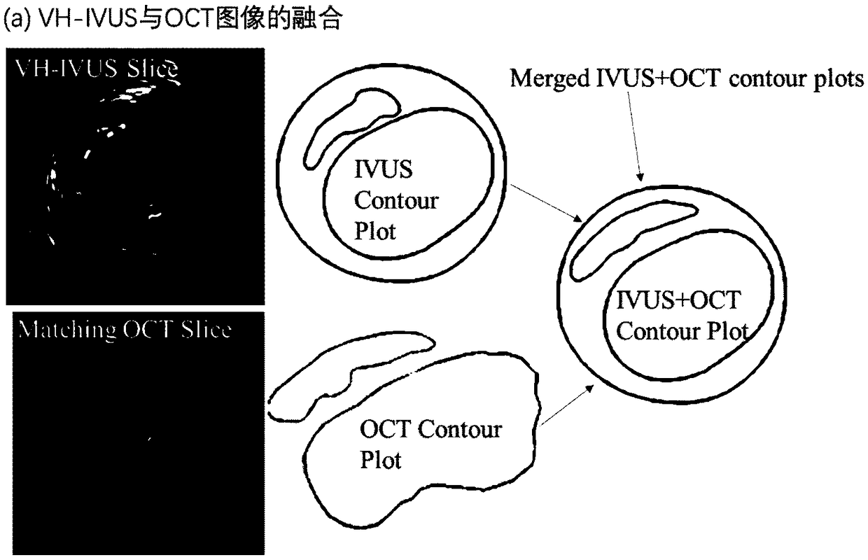
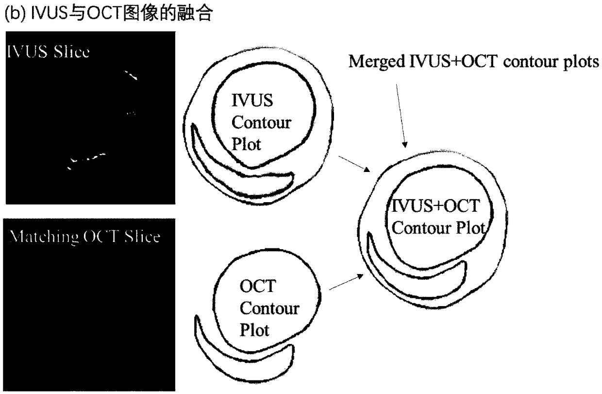
[0053] Step 2: After obtaining IVUS and OCT at T1 and T2 of the patient, adjust the IVUS image resolution to be consistent with the OCT image resolution, and then pair the images according to some characteristics of blood vessels and plaques as markers. Firstly, the IVUS and OCT images at T1 are paired, and the bifurcation of blood vessels is used as the first feature mark to determine the pairing of IVUS and OCT im...


PUM


Abstract

The invention belongs to the technical field of medical imaging, and particularly relates to a method for predicting the growth of coronary atherosclerotic plaque. The method comprises the steps thatIVUS images and OCT images of at least two moments are segmented respectively, then the segmented IVUS images and OCT images are fused, a three-dimensional multi-modality fluid-solid coupling model with cyclic bending is established according to a fused contour, a mechanical result of the model is calculated, risk factors of mechanics and morphology are extracted in combination with a morphological result, a plaque growth indicator is given, and the growth of the plaque is predicted by using a machine learning method, thereby providing an accurate and reliable method for predicting the growthof coronary plaque. The method for predicting the growth of the coronary atherosclerotic plaque provides the possibility of clinical application for plaque growth prediction.

Description

technical field [0001] The invention relates to a method for predicting the growth of coronary atherosclerotic plaque, belonging to the technical field of medical imaging. Background technique [0002] The prevalence and mortality rate of cardiovascular diseases in China are still on the rise. It is estimated that the number of patients with cardiovascular diseases is 290 million, and the death rate of cardiovascular diseases accounts for more than 40% of the residents' death from diseases, ranking first. For details, please refer to Chen Weiwei et al. Summary of China Cardiovascular Disease Report 2017, Chinese Journal of Circulation, 2018,33(01):1-8. The mechanisms leading to plaque rupture are unknown, and available screening and diagnostic methods are insufficient to identify victims before an event occurs. Current techniques for diagnosing coronary atherosclerosis often lack accurate and reliable computational mechanics analysis. Available medical imaging devices such...

Claims


Application Information

Patent Timeline
IPC IPC(8): A61B8/08A61B8/12A61B5/00
CPCA61B5/0066A61B8/0891A61B8/12A61B8/5246
Inventor 郭小亚唐达林曹进德
Owner SOUTHEAST UNIV
Why Eureka
Try Eureka