Exposure device and method for minimally invasive operating room septal defect

An operating room and spacer technology, which is applied in the field of medical devices, can solve the problems of difficulty in pulling and exposing the ventricular septal defect, restrictions on the popularization and application of minimally invasive cardiac surgery, narrow and long paths, etc., to reduce heart damage, facilitate operation, and simple operation Effect

Pending Publication Date: 2019-01-18
重庆医科大学附属第三医院 +1
View PDF0 Cites 2 Cited by
  • Summary
  • Abstract
  • Description
  • Claims
  • Application Information

AI Technical Summary

Problems solved by technology

[0003] At present, in minimally invasive ventricular septal defect repair surgery, nerve hooks are generally used to stretch and expose the ventricular septal defect; however, due to the sharp parts of the nerve hook, it is easy to cause heart damage during the process of pulling and exposing the ventricular septal defect In addition, due to the small incisions of various minimally invasive surgeries and the narrow and long path from the surgical field, the extension and removal of the nerve hook will be limited by the distance of the surgical site, resulting in difficulties in pulling and exposing the ventricular septal defect, which is limited to a certain extent. Promote the application of minimally invasive cardiac surgery

Method used


Image

  • Exposure device and method for minimally invasive operating room septal defect
  • Exposure device and method for minimally invasive operating room septal defect
  • Exposure device and method for minimally invasive operating room septal defect

Examples


Embodiment 1

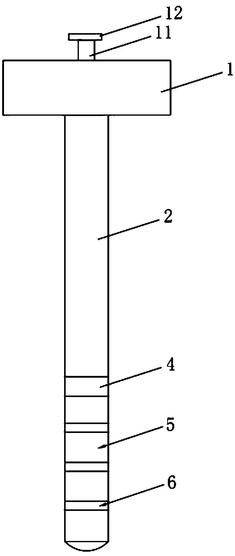
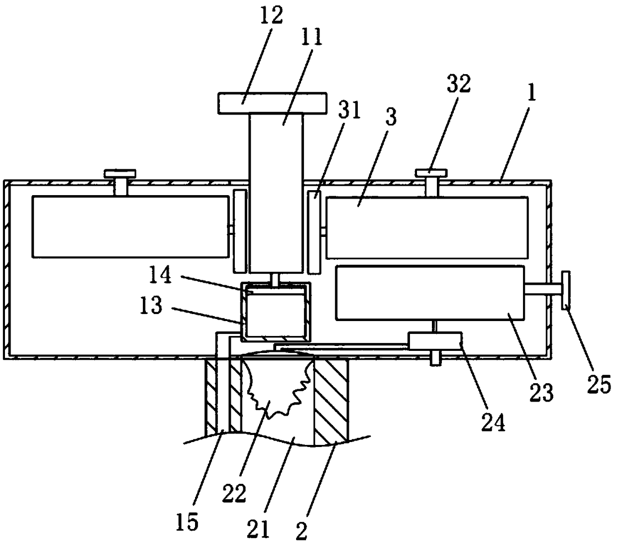
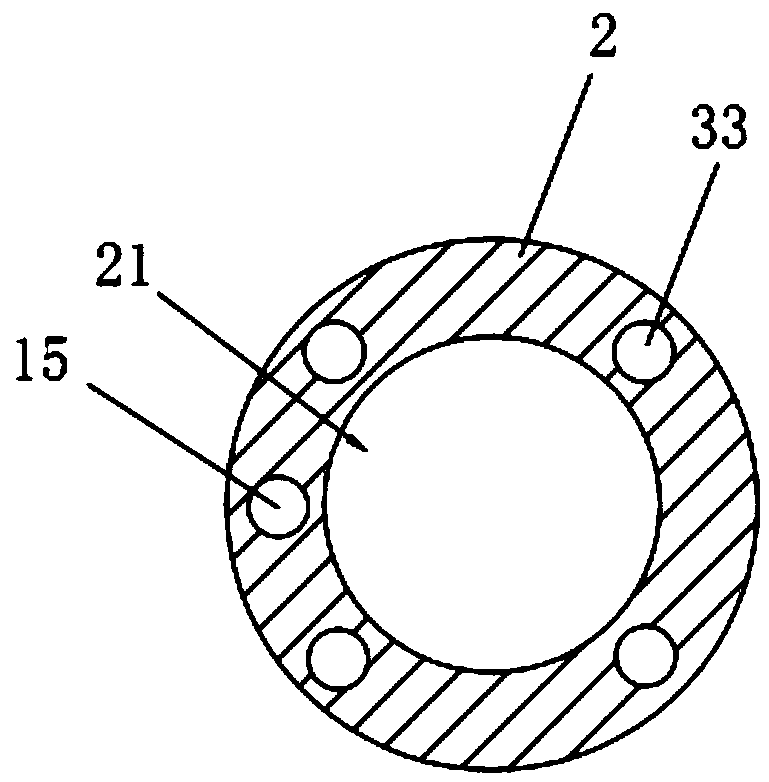
[0037] Embodiment 1: minimally invasive operating room septal defect exposure device, such as figure 1 and figure 2 As shown, it includes a hand-held box 1 , a tube body 2 fixedly connected to the hand-held box 1 and a first airbag 4 fixedly connected to the outer wall of the tube body 2 , and the first airbag 4 is arranged along the circumferential direction of the tube body 2 . The hand-held box 1 is provided with a syringe 13 for storing physiological saline, and the tube body 2 is provided with an infusion tube 15 with two ends communicating with the syringe 13 and the first air bag 4 respectively. A sealing piston 14 is movably connected to the syringe 13 , and an operating rod 11 for driving the sealing piston 14 to move is movably connected to the hand-held box 1 . Utilizing the first airbag 4, in the process of pulling and exposing the ventricular septal defect, the tube body 2 and the ventricular septal defect are prevented from being detached from each other. Util...

Embodiment 2

[0044] Embodiment 2: minimally invasive operation room septal defect exposure method, such as figure 2 and Figure 5 shown, including the following steps:

[0045] Step 1, the tube body 2 is sequentially passed through the surgical incision, chest cavity, right atrium, tricuspid valve, right ventricle, ventricular septum and left ventricle until the first air bag 4 on the tube body 2 is completely placed in the left ventricle.

[0046] Step 2, then press the operating lever 11, and the physiological saline in the syringe 13 is delivered into the first airbag 4 through the infusion pipeline 15 under the extrusion of the sealing piston 14 until the inside of the first airbag 4 is filled with physiological saline.

[0047] Step 3, after pulling the tube body 2, the first air bag 4 touches the inner wall of the left ventricle, and the tube body 2 exposes the ventricular septum during the pulling process.

[0048] Such as figure 2 and Figure 4 As shown, in the process of ins...


PUM


Abstract

The invention discloses an exposure device and a method for minimally invasive operating room septal defects, and relates to the technical field of medical apparatus and instruments. The invention solves the cardiac injury caused easily in the process of stretching and exposing the ventricular septal defect, The technical scheme comprises a hand-held box, a tube fixedly connected with the hand-held box and a first air bag fixedly connected with the outer wall of the tube, wherein the first air bag is circumferentially arranged along the tube body; The hand-held box is provided with a syringe for storing normal saline, and the pipe is provided with a liquid infusion pipe which is respectively communicated with the syringe and the first air bag at two ends. The injection barrel is movably connected with a sealed piston, and the hand box is movably connected with an operation rod for driving the sealed piston to move, which has the effects of reducing heart injury and simplifying the operation of pulling and exposing the ventricular septal defect.

Description

technical field [0001] The invention relates to the technical field of medical instruments, more specifically, it relates to a minimally invasive operating room septal defect exposure device and method. Background technique [0002] Ventricular septal defect is the most common congenital cardiac malformation clinically. It can exist alone or in combination with other cardiac malformations. The main reason is that the ventricular septum is underdeveloped in the embryo, forming abnormal communication, and generating abnormal shunt between the left and right ventricles. [0003] At present, in minimally invasive ventricular septal defect repair surgery, nerve hooks are generally used to stretch and expose the ventricular septal defect; however, due to the sharp parts of the nerve hook, it is easy to cause heart damage during the process of pulling and exposing the ventricular septal defect In addition, due to the small incisions of various minimally invasive surgeries and the...

Claims


Application Information

Patent Timeline
Patent Type & Authority Applications(China)
IPC IPC(8): A61B17/02A61B17/00
CPCA61B17/0218A61B17/00234A61B2017/00243A61B2017/0237
Inventor 李福平高凌云林德清陈明祥唐文帅胡文平张为民李志平黎小平
Owner 重庆医科大学附属第三医院
Why Eureka
Try Eureka