A medical image segmentation method based on a one-way multi-task convolution neural network

A technology of convolutional neural network and medical image, which is applied in the field of medical image segmentation based on single-pass multi-task convolutional neural network, can solve the problems of complex model structure, very high hardware configuration requirements, and huge parameter volume, so as to improve the complexity of the system. degree, alleviate the problem of category imbalance, and reduce the effect of system complexity

Inactive Publication Date: 2019-02-19
SOUTH CHINA UNIV OF TECH
View PDF4 Cites 17 Cited by
  • Summary
  • Abstract
  • Description
  • Claims
  • Application Information

AI Technical Summary

Problems solved by technology

However, the model structure used in deep learning is often very complex, and an extremely large amount of p...

Method used


Image

  • A medical image segmentation method based on a one-way multi-task convolution neural network
  • A medical image segmentation method based on a one-way multi-task convolution neural network
  • A medical image segmentation method based on a one-way multi-task convolution neural network

Examples


Embodiment

[0032] In order to describe the content of this embodiment conveniently, first, a brief description of the English abbreviations and technical terms in this embodiment is given:

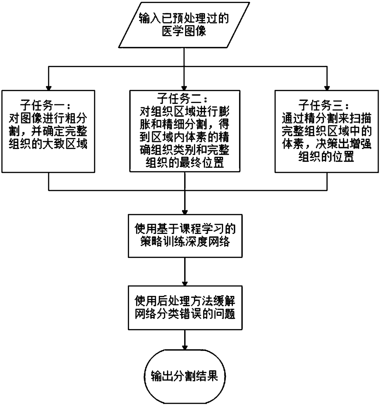
[0033] Tissue classification and segmentation tasks: In the medical field, there are four classifications for the variant tissue of the brain region, namely: edematous tissue, necrotic tissue, non-enhanced tissue, and enhanced tissue; these four types of tissue are classified into Three overlapping areas, including: full tissue, tissue core, and enhanced tissue.

[0034] Flair sequence: One of the sequences of magnetic resonance images, commonly known as pressurized water images. In this sequence, the gray matter is hyperintense (the image appears "brighter") and the white matter is hypointense (the image appears "darker"), and these properties are similar to T2WI. But on the pressurized water image, the cerebrospinal fluid is low signal, that is, black.

[0035] Ground-truth: In foreign scientific...


PUM


Abstract

The invention discloses a medical image segmentation method based on a one-way multi-task convolution neural network. Firstly, the segmentation task is divided into three different but related sub-tasks by using the network. A first sub-task carries out rough segmentation on that image to determine a rough area of the whole organization; a second subtask dilates and fine-segments the area to getthe exact tissue type and exact position of the whole tissue. Subtask 3 uses refined segmentation to scan voxels in the intact tissue and determine the location of the enhanced tissue. Then the threesub-tasks are trained in stages with course learning strategy from easy to difficult to realize data and parameter sharing among sub-tasks. Finally, the post-processing method is used to improve the classification error problem and output the final segmentation results. The invention synthesizes the associated sub-tasks into a single network for simultaneous training, improves the shortcomings ofcascade model training sub-tasks one by one and better solves the category imbalance problem, realizes the segmentation of the image from coarse to fine by one-way operation, and improves the segmentation effect while reducing the complexity of the system.

Description

technical field [0001] The invention relates to the field of image processing, in particular to a medical image segmentation method based on a single-pass multi-task convolutional neural network. Background technique [0002] With the development of science and technology, modern medical treatment such as computed tomography (CT), magnetic resonance imaging (Magnetic Resonance Imaging, MRI), positron emission tomography (PET) and electronic endoscopy (Endoscopy) Imaging technology and equipment have been rapidly developed and popularized. After these technologies and equipment are put into use, we can easily obtain a large amount of medical imaging data, and process, observe, and analyze the patient's diseased tissues or organs through computers and other equipment, so that medical personnel can understand the condition more clearly and greatly improve the quality of life. Shorten the diagnosis time and improve the diagnostic accuracy. It is worth noting that medical image...

Claims


Application Information

Patent Timeline
IPC IPC(8): G06T7/11G06K9/62
CPCG06T7/11G06T2207/10088G06T2207/30016G06T2207/20084G06T2207/20081G06F18/23213G06F18/241
Inventor 丁长兴周晨红黄英杰
Owner SOUTH CHINA UNIV OF TECH
Why Eureka
Try Eureka