

Use of umbilical cord blood derived exosomes for tissue repair
A technology of exosomes and umbilical cord blood, which can be applied in the fields of mammalian medical raw materials, medical preparations containing active ingredients, and drug combinations, etc., which can solve the problems of unassessed biological activity and unassessed chronic wound treatment potential.
- Summary
- Abstract
- Description
- Claims
- Application Information
AI Technical Summary
Problems solved by technology
Method used
Image

Examples
Embodiment 1
[0194] Example 1. Ischemic preconditioning stimulates umbilical cord blood mononuclear cells (UCBMNC) to secrete exosomes and increase their biological activity on skin cells.
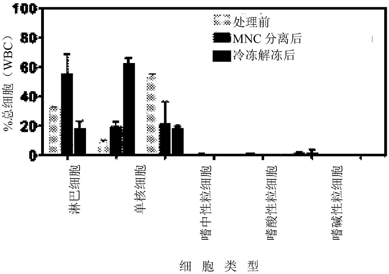
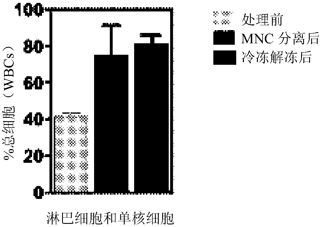
[0195] Using Lymphoprep TM The total fraction of UCBMNCs was isolated and the cells were characterized using a hematology analyzer. After cryopreservation, the population was enriched in lymphocytes and monocytes (MNC), while most of the neutrophils were lost ( Figure 1A with Figure 1B ). The pool used to secrete exosomes is mainly composed of lymphocytes and monocytes.
[0196] Experiments show that ischemic preconditioning (0.5% O 2 , 5% CO 2 , 0% FBS, 18h) stimulated UCBMNCs to secrete bioactive exosomes, which were internalized with specific kinetics by different types of skin cells (fibroblasts, keratinocytes and endothelial cells). Experiments also demonstrated that these exosomes (UCBMNC-Exo) exert potent pro-regenerative effects in vitro, which are involved in increasing the survival and...
Embodiment 11
[0197] Example 1.1: Characterization of exosomes
[0198] Experimental procedure:
[0199] Exosomes were purified by differential centrifugation as described by Thery C. (2006) et al., which is incorporated herein in its entirety.
[0200] Briefly, the supernatant was subjected to two successive centrifugation steps at 300 g (10 min), 2000 g (20 min) and 10,000 g (30 min) to remove cells, cell debris and microvesicles, respectively. After centrifugation at 100,000g for 2 hours in a SW41Ti or SW32Ti floating bucket rotor (Beckman), the pellet was washed with 7 mL of PBS and centrifuged for an additional 2 hours at 100,000g. Resuspend the exosome-containing pellet in 250 µL of PBS. Using from BioRad TM The DC Protein Assay Kit quantifies exosomes based on their protein content.
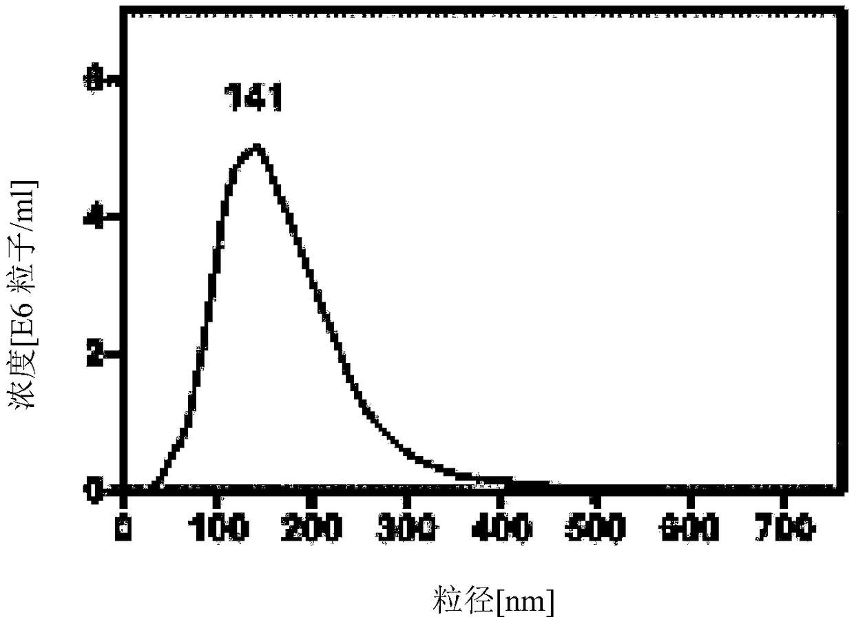
[0201] Exosomes secreted by UCBMNCs were initially characterized using transmission electron microscopy (TEM) and dynamic light scattering (DLS). DLS analysis was performed on a ζPALS zeta potentia...
Embodiment 12
[0205] Example 1.2: Kinetics of exosome uptake by skin cells
[0206] 1.2.1 Percentage of skin cells
[0207] Experimental procedure:
[0208] Skin cells uptake UCBMNC-derived exosomes, however, uptake kinetics and magnitude depend on cell type. To demonstrate differences in the kinetics of exosome uptake by skin cells, exosomes were initially labeled with fluorescent dyes (NBD-DPPE and Syto green). Briefly, NBD-DPPE (16 μM in ethanol) or Syto Green (10 μM in DMSO) dyes were added to exosome suspensions (100 μL; protein concentrations ranged from 60 to 70 μg / mL) (DMSO and EtOH< 1%) and incubate at 37°C for 30 minutes. After blocking the reaction with bovine serum albumin (BSA, 1%, w / v), the exosomes were washed with PBS (6 mL), and free dye was removed by ultracentrifugation at 100,000 g for 1 hour. Then, the exosomes were resuspended in PBS (100 μl). The procedure is always performed with fresh exosomes. Labeled exosomes were used immediately for internalization experim...
PUM
| Property | Measurement | Unit |
|---|---|---|
| particle size | aaaaa | aaaaa |
| concentration | aaaaa | aaaaa |
| particle size | aaaaa | aaaaa |
Abstract
Description
Claims
Application Information