La/single-stranded binding protein (La/SSB) chimera antigen modified NK cell, and preparation method and application thereof
A technology of NK cells and host cells, applied in genetically modified cells, cells modified by introducing foreign genetic material, blood/immune system cells, etc. disorder, etc.
- Summary
- Abstract
- Description
- Claims
- Application Information
AI Technical Summary
Problems solved by technology
Method used
Image
Examples
Embodiment 1
[0203] Embodiment 1 Purification of LaA protein and LaA-BCR protein
[0204] 1.1 Induction and purification of LaA protein
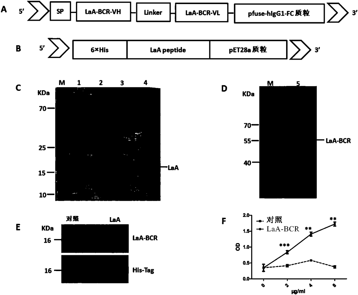
[0205] To induce the expression of soluble LaA protein, the cDNA encoding LaA protein was subcloned into pET28a prokaryotic expression vector (LaA-pET28a) containing 6×HIS ( figure 1 B), 6×HIS tag is used for protein purification. Then the LaA-pET28a plasmid was transformed into E.coli BL21(DE3) strain for culture.
[0206] The LaA protein was induced and purified according to the following steps: Pick a single LaA-pET28a clone and culture it overnight at 37°C in 3 ml LB medium containing 100 μg / mL kanamycin. Then continue to cultivate in the fresh culture medium that contains 100 μ g / mL kanamycin according to the ratio dilution of 1:100, when bacterium early logarithmic phase (OD=0.6-0.8), add 0.5mMisopropylβ-D-1-thiogalactopyranoside ( IPTG, Sangon Biotech) at 37°C for 2.5-3 hours. Then LaA protein was purified by Ni-NTA agarose (GE Healthcare Life...
Embodiment 2
[0211] Example 2 Affinity Determination of LaA-BCR Protein and LaA Antigen
[0212] Purified LaA antigen added to 5×SDS-PAGE loading buffer (P0015L, Beyotime, Shanghai, China) and non-LaA protein as a negative control were used as samples, separated by 12% SDS-PAGE, and the proteins on SDS-PAGE were separated by Transfer to PDVF membrane (IPVH00010, Millipore, MA), block with 5% skimmed milk at room temperature for 1 hour (Guangming, Shanghai, China), apply purified LaA-BCR protein and incubate overnight at 4°C, and then apply 0.1% Tween- After washing three times with 20 PBST, the PVDF membrane was incubated with horseradish peroxidase-conjugated anti-mouse IgG (H+L) secondary antibody (A10677, Life Technologies, CA). After washing three times with PBST, the PDVF membrane was visualized by chemiluminescence by applying the enhanced chemiluminescence kit (WBKLS0500, Millipore Corporation, MA).
[0213] Purified LaA protein was coated on ELISA microplate overnight at 4°C. The...
Embodiment 3
[0215] Example 3 Construction, characteristics and functional verification of LaA-CAAR NK92MI cells
[0216] 3.1 Structure design and lentiviral transfection of LaA-CAAR
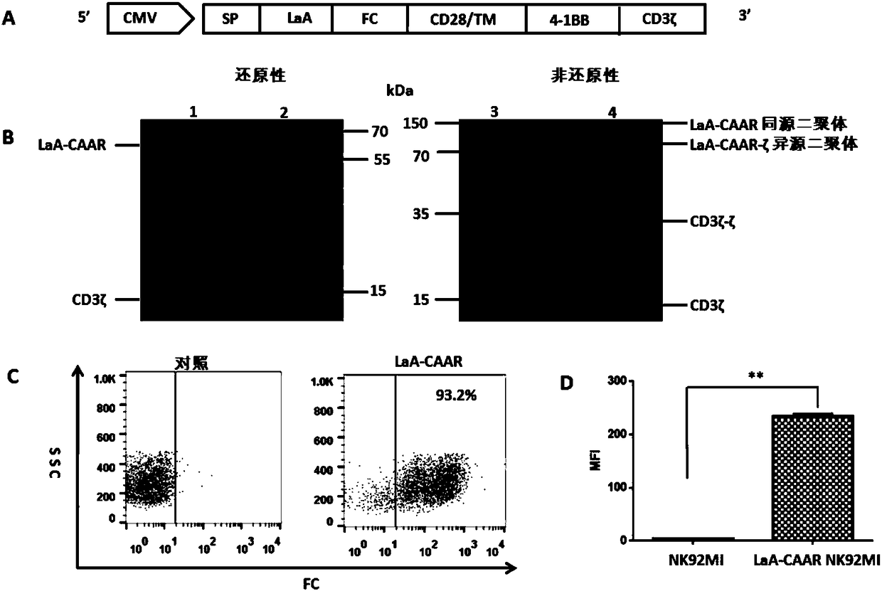
[0217] The molecular structure design of LaA-CAAR includes: a CD8 transmembrane signal peptide guides the expression of LaA-CAAR on the surface of NK92MI cells, the LaA antigen fragment is used as an extracellular fragment, F C Linking fragments, CD28 molecule (comprising transmembrane region), 4-1BB co-stimulatory factor and CD3ζ signal region are connected to form a fusion protein ( figure 2 A). The LaA-CAAR fusion protein contains: leader sequence, LaA peptide, hinge domain (FC), CD28 transmembrane domain TM, two co-stimulatory domains (CD28 and 4-1BB) and CD3ζ intracellular signaling domain.
[0218] The SP-LaA-Fc-CD28 / TM-4-1BB-CD3ζ fusion gene sequence was gene-synthesized, and then constructed into the pCDH-CMV-MCS-EF1-CopPuro (System Biosciences) lentiviral expression vector. Plate 293T cells, pac...
PUM
 Abstract
Description
Claims
Application Information
 - R&D
- Intellectual Property
- Life Sciences
- Materials
- Tech Scout
- Unparalleled Data Quality
- Higher Quality Content
- 60% Fewer Hallucinations
Browse by: Latest US Patents, China's latest patents, Technical Efficacy Thesaurus, Application Domain, Technology Topic, Popular Technical Reports.
© 2025 PatSnap. All rights reserved.Legal|Privacy policy|Modern Slavery Act Transparency Statement|Sitemap|About US| Contact US: help@patsnap.com


