
Spinal column pressurizing device used for nuclear magnetic resonance imaging

A technology of nuclear magnetic resonance imaging and pressure device, applied in the field of biomedical engineering

Active Publication Date: 2019-03-08
CAPITAL UNIVERSITY OF MEDICAL SCIENCES
View PDF20 Cites 1 Cited by
  • Summary
  • Abstract
  • Description
  • Claims
  • Application Information

AI Technical Summary

Problems solved by technology

At present, similar pressurization devices are attached with metal materials, and the pressurization can only be completed before the inspection, and the metal parts are removed during the inspection, so real-time pressurization adjustment and pressure measurement on the MRI bed cannot be realized.

Method used


Image

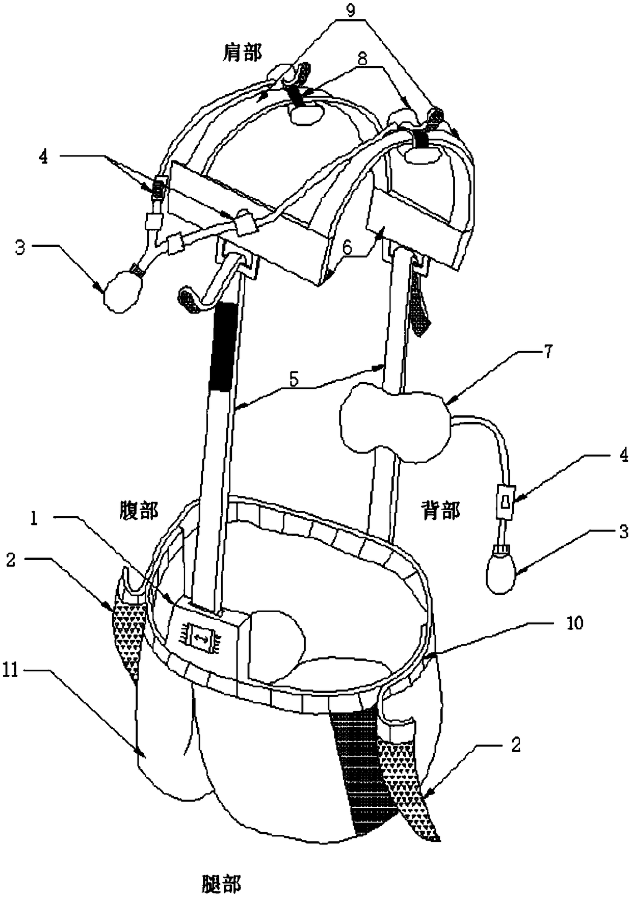
  • Spinal column pressurizing device used for nuclear magnetic resonance imaging

Examples


Embodiment Construction

[0015] In order to further understand the present invention, preferred solutions of the present invention will be described below in conjunction with examples. These descriptions only illustrate the features and advantages of the present invention, but do not limit the protection scope of the present invention.

[0016] Such as figure 1 Shown is a schematic diagram of a spinal compression device for magnetic resonance imaging, the device consists of a load measuring instrument 1, a leg Velcro 2, an air inflation device 3, a ventilation valve 4, a connecting belt 5, a connecting beam 6, and a waist airbag 7. The shoulder airbag 8, the shoulder belt 9, the waist belt 10, and the detachable shorts 11 form. The shoulder airbag 8 is worn on the shoulder strap 9 and can slide forward and backward on the shoulder strap 9 so as to adjust the position of the shoulder airbag 8 . There is a connecting crossbeam 6 on the chest and the back respectively, and the shoulder belt 9 is connec...


PUM


Abstract

The invention discloses a spinal column pressurizing device used for nuclear magnetic resonance imaging. The device structurally comprises components of detachable shorts, connecting belts, shoulder straps, a waist belt, shoulder spherical bags, a waist spherical bag, a load measuring instrument, a connecting hook-and-loop fastener, leg hook-and-loop fasteners and the like; non-metal materials areadopted by the components, and therefore the components have no influence on the nuclear magnetic resonance imaging. The detachable shorts are connected with the shoulder straps through the connecting belts, the connecting belts can be bonded at different positions through a hook-and-loop fastener connecting manner, and people with different body shapes can conveniently worn and adapt. The connecting belts are provided with pulling force load measuring instruments, and the pressurized magnitude on a spinal column can be displayed in real time by adopting an indicator and a ruler. Pressurizingair bags are arranged on the shoulders and the waist so that the pressurized magnitude can be adjusted at will when nuclear magnetic resonance inspection is conducted, and the force bearing states inwhich a tested person feels uncomfortable of painful when bearing loads are simulated. The pressurizing device has the advantages that the pressurizing device can be adapted to tested people with different heights and shapes, and the free adjusting of the pressurizing magnitude on the spinal column performed by the tested person when the nuclear magnetic resonance inspection is conducted is convenient.

Description

technical field [0001] The present invention relates to the field of biomedical engineering. In particular, the invention relates to a spinal compression device for magnetic resonance imaging, which can be worn by a subject during magnetic resonance imaging and can apply different pressures to the spine so that the subject is under load. An MRI of the lower spine. Background technique [0002] The human spine is an important load-bearing structure. Each segment must carry a large amount of load and perform complex and diverse movements, which is very prone to damage and degeneration. Whether it is the herniation of the intervertebral disc caused by the accelerated pace of work, or the fracture and fracture of the spine caused by external force, if it is not treated in time, it will cause irreversible consequences. [0003] At present, MRI is an important means of examining spinal injuries, but there are also some limitations. For example, no injury is found when the subjec...

Claims


Application Information

Patent Timeline
Patent Type & Authority Applications(China)
IPC IPC(8): A61B5/055
CPCA61B5/055
Inventor 宋红芳刘志成赵志强吴宝平李琦龙晓雪
Owner CAPITAL UNIVERSITY OF MEDICAL SCIENCES
Learn More