
Fixing device for super-resolution imaging of red blood cell and use method

A technology of super-resolution imaging and fixing devices, which is applied in the directions of measuring devices, analyzing materials, and analyzing materials through optical means. Effect

Pending Publication Date: 2019-04-05
DALIAN UNIV OF TECH
View PDF5 Cites 0 Cited by
  • Summary
  • Abstract
  • Description
  • Claims
  • Application Information

AI Technical Summary

Problems solved by technology

However, the current super-resolution imaging of single-molecule localization is mostly based on total internal reflection technology. This technology requires the observed biological samples to be observed close to the surface of the glass slide. Observation of cells is difficult to achieve

Method used


Image

  • Fixing device for super-resolution imaging of red blood cell and use method
  • Fixing device for super-resolution imaging of red blood cell and use method
  • Fixing device for super-resolution imaging of red blood cell and use method

Examples


Embodiment 1

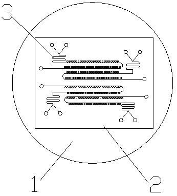
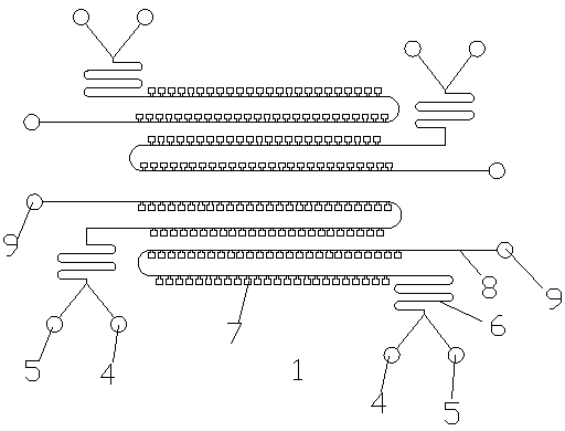
[0028] Figure 1-Figure 4 Structural diagram of the fixture for super-resolution imaging of red blood cells. There are four microchannels 3 in the fixture, indicated in red and blue respectively, and each channel has 90 red blood cell fixation chamber repeating units. The thickness of each red blood cell fixation chamber is 1um, and the chamber size is 50um*50um. The experimental process is as follows. The PDMS microchamber structure is prepared by soft lithography and drilling. The PDMS microchamber structure is punched down on the clean glass slide after plasma treatment, and the blood sample is evenly shaken and then passed into the sample inlet 4. , due to the wettability of the PDMS microchamber 2 after plasma treatment and the characteristics of gas absorption, the blood sample enters the inlet main channel 6 from the sample inlet 4 and communicates with the fixed chamber main channel 7b, and then passes through the fixed chamber inlet 7c communicates with the erythroc...

Embodiment 2

[0030] Figure 5 Super-resolution imaging of adult erythrocytes using the fixation device proposed by the present invention. Among them, Figure a is 5 red blood cells in ordinary wide-field imaging, Figure b is 5 red blood cells in super-resolution imaging, Figure c indicates that the positioning accuracy of super-resolution imaging is 20nm, Figure d shows that the chip can keep red blood cells still within 80s, and carry out For super-resolution imaging, both dyes dii and c180 can be realized. Scale bar 2um.

Embodiment 3

[0032] This fixture can be used for super-resolution imaging of red blood cells in young children with pneumonia

[0033] (1) Prepare the PDMS microchamber structure by soft lithography and drilling;

[0034] (2) After the PDMS microchamber structure is treated by plasma, the graphic surface is washed down and placed on a clean glass slide;

[0035] (3) The blood of children with pneumonia was oscillated evenly and passed into the chip. Due to the wettability of PDMS to liquid and the characteristics of gas absorption after plasma treatment, the blood was automatically absorbed into the red blood cell fixation chamber;

[0036] (4) Since the height of the red blood cell fixation chamber does not exceed twice the length of the red blood cell line, the red blood cells are fixed in a single layer after being sucked into the red blood cell fixation chamber;

[0037] (5) The microfluidic chip loaded with red blood cells is placed on a super-resolution microscope for imaging observ...


PUM

PropertyMeasurementUnit
Thicknessaaaaaaaaaa
Thicknessaaaaaaaaaa

Abstract

The invention discloses a fixing device for super-resolution imaging of a red blood cell and a use method, and belongs to the technical field of interaction of micromachining technology and biologicalimaging. According to the fixing device for super-resolution imaging of the red blood cell, the red blood cell is fixed to and attached to the surface of glass, physical fixation of the red blood cell with a smooth surface is realized, total internal reflection super-resolution imaging observation is convenient, and the requirements for sample wall-attaching in the total internal reflection super-resolution imaging process are met. The device does not need to be chemically modified or processed, the use is simple and convenient, and the manufacturing cost is low. The size of each red blood cell fixing chamber is small, a plurality of repetitive units can be arranged on the fixing device with the area less than the area of a one-yuan coin, a plurality of red blood cells can be subjected tosuper-resolution shooting synchronously, in addition, a plurality of micro-channels can be arranged on one fixing device, that is, red blood cells in blood samples of different sources can be subjected to super-resolution imaging synchronously on one fixing device, and the experiment throughput is greatly improved.

Description

technical field [0001] The invention relates to a fixing device and a using method for super-resolution imaging of red blood cells, which belong to the technical field intersecting micro-processing technology and biological imaging. Background technique [0002] Super-resolution imaging technology is a field of rapid development in recent years. Because it can break through the optical diffraction limit and observe biological samples at the nanometer scale, it has now become a powerful tool for cell analysis. However, the current super-resolution imaging of single-molecule localization is mostly based on total internal reflection technology. This technology requires the observed biological samples to be observed close to the surface of the glass slide. Observation of cells is difficult to achieve. [0003] The fixing device for super-resolution imaging of red blood cells proposed by the present invention uses elastic material PDMS to fix red blood cells in a chamber, and th...

Claims


Application Information

Patent Timeline
IPC IPC(8): G01N21/01G01N21/84
CPCG01N21/01G01N21/84
Inventor 杨薇王淑静叶智伟
Owner DALIAN UNIV OF TECH
Learn More