
Chest focus image classification method and device, computer equipment and storage medium

A classification method and lesion technology, applied in image analysis, computer parts, computing, etc., can solve the problems of high misdiagnosis rate, shortage of social medical resources, time-consuming and labor-intensive, etc., so as to improve the accuracy rate and alleviate the shortage of social medical resources. , the effect of improving the efficiency of diagnosis

Pending Publication Date: 2019-04-26
PING AN TECH (SHENZHEN) CO LTD
View PDF0 Cites 15 Cited by
  • Summary
  • Abstract
  • Description
  • Claims
  • Application Information

AI Technical Summary

Problems solved by technology

Limited by the medical experience and knowledge of the existing medical staff, there will be great differences in the judgment of the disease, and in the absence of consultation, the diagnostic results obtained by the medical staff based on their experience cannot be verified by other personnel and questioning, it often occurs that the best treatment time of the patient is delayed due to misdiagnosis, and even the death of the patient is caused.
[0004] Therefore, the chest X-ray disease diagnosis in the prior art only relies on the personal experience of medical staff, which will lead to a higher misdiagnosis rate and endanger the life and health of patients.
At the same time, due to human judgment, the diagnosis process is time-consuming, labor-intensive, and inefficient, resulting in a shortage of social medical resources.

Method used


Image

  • Chest focus image classification method and device, computer equipment and storage medium
  • Chest focus image classification method and device, computer equipment and storage medium
  • Chest focus image classification method and device, computer equipment and storage medium

Examples


Embodiment Construction

[0073] In order to enable those skilled in the art to better understand the solutions of the present invention, the technical solutions in the embodiments of the present invention will be clearly and completely described below in conjunction with the drawings in the embodiments of the present invention.

[0074] In some processes described in the specification and claims of the present invention and the above-mentioned drawings, a plurality of operations appearing in a specific order are contained, but it should be clearly understood that these operations may not be performed in the order in which they appear herein Execution or parallel execution, the serial numbers of the operations, such as 101, 102, etc., are only used to distinguish different operations, and the serial numbers themselves do not represent any execution order. Additionally, these processes can include more or fewer operations, and these operations can be performed sequentially or in parallel. It should be n...


PUM


Abstract

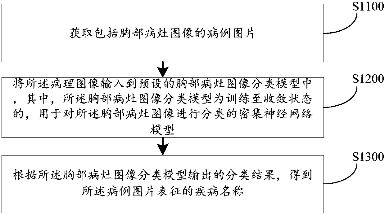
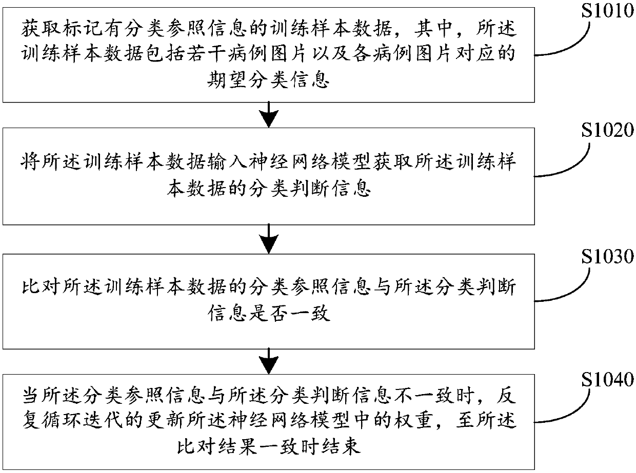
The embodiment of the invention discloses a chest lesion image classification method and device, computer equipment and a storage medium. The method comprises the steps that a case image comprising achest lesion image is acquired; Inputting the case image into a preset chest focus image classification model; And obtaining a disease name represented by the case image according to a classificationresult output by the chest focus image classification model. When the chest focus image classification model is trained, a confirmed or cured disease picture is adopted, so that the chest focus imageclassification model has the capability of judging the disease name in the same type of picture, and the disease judgment represented by the chest focus image classification model on the image can represent the common judgment result in the field. Therefore, by means of the chest focus image classification model, the limitation that judgment only depends on experience of a single medical worker ora part of medical workers during disease diagnosis can be avoided, the judgment result of the disease conforms to the general recognition standard in the field, and the disease diagnosis accuracy canbe effectively improved.

Description

technical field [0001] Embodiments of the present invention relate to the field of model algorithms, in particular, a method, device, computer equipment, and storage medium for classifying images of chest lesions. Background technique [0002] At present, chest X-ray examination is the most common method for diagnosing chest diseases, and is often used to check diseases of the thorax (including ribs, thoracic spine, soft tissue, etc.), chest cavity, lung tissue, mediastinum, heart, etc. Such as pneumonia, tumor, fracture, pneumothorax, cor pulmonale, heart disease. [0003] In the prior art, the chest X-ray examination can only generate the patient's chest X-ray, and the medical staff can diagnose the patient's disease by observing whether there is a lesion image in the chest X-ray, or observing the position and shape of the lesion image. That is to say, in the prior art, the diagnosis of the user's disease can only be judged by the individual medical staff. Limited by the...

Claims


Application Information

Patent Timeline
IPC IPC(8): G06K9/62G06T7/00
CPCG06T7/0012G06T2207/30096G06T2207/20084G06T2207/20081G06T2207/10081G06V2201/032G06F18/24G06F18/214
Inventor 刘新卉马进王健宗肖京
Owner PING AN TECH (SHENZHEN) CO LTD
Learn More