Output method and device in lung segment segmentation of CT image

A CT image and output method technology, applied in image analysis, image enhancement, image data processing, etc., can solve problems such as poor segmentation effect and uncertain detection results, and achieve high accuracy, improved diagnosis rate, and efficient control.

Inactive Publication Date: 2019-04-26
HANGZHOU YITU MEDIAL TECH CO LTD
View PDF4 Cites 7 Cited by
  • Summary
  • Abstract
  • Description
  • Claims
  • Application Information

AI Technical Summary

Problems solved by technology

This method ignores the differences in lung structure between different patients, and the detection results are uncertain (I.C. Sluimer, etc.: "Towards automated segmentation of the pathological lung in CT," IEEE Trans.Med.Imag., vol.24 , no.8, pp.1025–1038, Aug.2005.)
2) Establish a water-washing deformation through the analysis of the trachea and vessels. This method has a better segmentation effect for visible fissures, but poor segmentation for partially invisible segments (E.van Rikxoort, etc.: "Automatic segmentation of pulmonary lobes robust against incomplete fissures." IEEE Transactions on Medical Imaging, vol.29, no.6, pp.1286–1296, 2010.)
[0005] Although the above patent improves the accuracy of lung nodule edge detection and segmentation in CT images, before starting to detect the edge of the image, it needs to be compared with the initially set threshold, and the detection can only be started when the conditions are met , on the contrary, the image needs to be reprocessed, the scope of this method is limited by individual differences, and continuous adjustment is required

Method used


Image

  • Output method and device in lung segment segmentation of CT image
  • Output method and device in lung segment segmentation of CT image
  • Output method and device in lung segment segmentation of CT image

Examples


Embodiment Construction

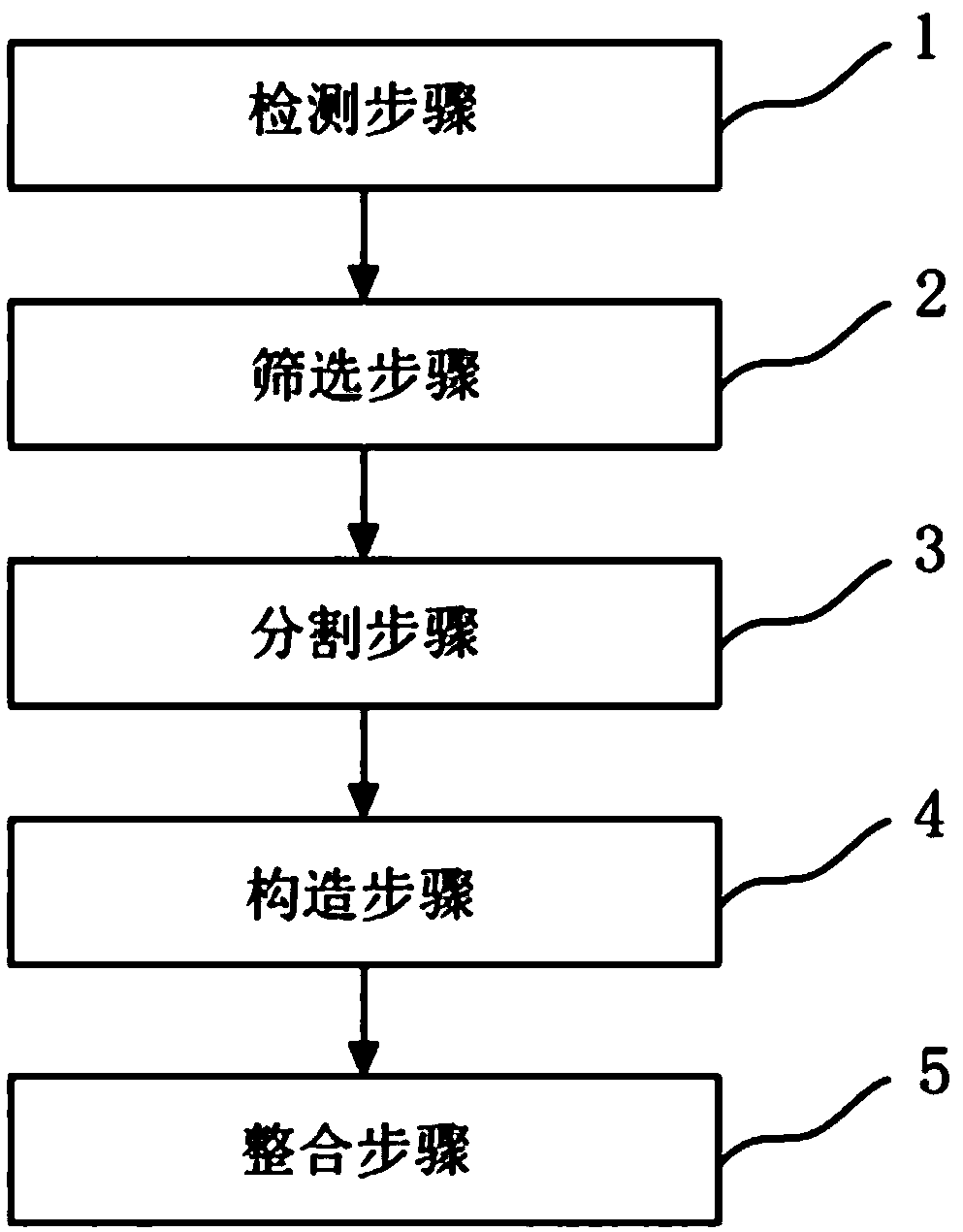
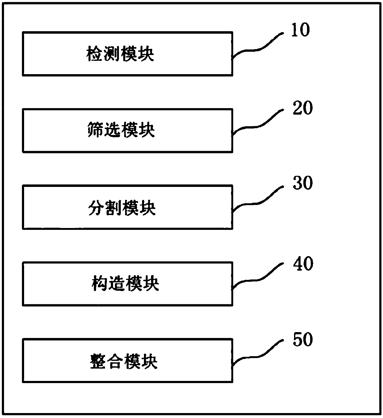
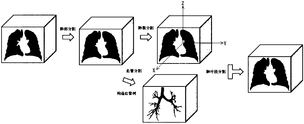
[0046] The implementation of the present invention will be illustrated by specific specific examples below, and those skilled in the art can easily understand other advantages and effects of the present invention from the content disclosed in this specification. Although the description of the present invention will be presented in conjunction with a preferred embodiment, it does not mean that the features of the invention are limited to this embodiment. On the contrary, the purpose of introducing the invention in conjunction with the embodiments is to cover other options or modifications that may be extended from the claims of the present invention. The following description contains numerous specific details in order to provide a thorough understanding of the present invention. The invention may also be practiced without these details. Also, some specific details will be omitted from the description in order to avoid obscuring or obscuring the gist of the present invention....


PUM


Abstract

The invention relates to a CT image lung lobe segment segmentation method, device and system, a storage medium and equipment, and the method comprises the steps: a detection step: in a CT image, detecting and outputting a lung contour which comprises an intra-lung region and an extra-lung region; A screening step: in the lung contour, selecting a machine segmentation mode to screen out an intra-lung region, and taking the intra-lung region as a candidate region; A segmentation step: in the 3D level of the candidate region, performing blood vessel segmentation and pulmonary lobe segmentation onthe lung segment and the pulmonary lobe at the same time; A construction step: according to a blood vessel segmentation result, constructing a blood vessel tree to obtain three-dimensional blood vessel distribution of the lung; And an integration step: combining the blood vessel tree with the lung split segmentation result, and carrying out lung segment segmentation to obtain a final segmentationresult of the candidate region. According to the CT image lung lobe segment segmentation method, device and system based on deep learning, the storage medium and the equipment, errors are effectivelyreduced, the diagnosis rate and accuracy are improved, and the CT image lung lobe segment segmentation method, device and system are not limited by individual lung morphological differences.

Description

technical field [0001] The present invention relates to the field of image segmentation, in particular to a method, device, system, storage medium and equipment for lung lobe segment segmentation of CT images. Background technique [0002] Lung cancer is currently the cancer with the highest mortality rate among all cancers. Pulmonary nodules are the image manifestations of lung cancer. They appear as shadows with increased density in CT imaging. The detection and segmentation of lung nodules in lung lobes and lung segments in CT images is of great importance for early screening and evaluation of lung cancer. significance. In existing detection, 3D CT (Emmenlauer, M., etc.: "free, fast and reliable stitching of large 3d datasets." J Microscopy, 233, no.1, pp.42-60, 2009.) image data volume Large, and there are large individual differences, which brings certain difficulties to the segmentation technology of lung lobes and lung segments. [0003] Different from the applicat...

Claims


Application Information

Patent Timeline
IPC IPC(8): G06T7/00G06T7/12G06T7/187G06K9/62
CPCG06T7/0012G06T7/12G06T7/187G06T2207/20084G06T2207/20081G06T2207/10081G06T2207/30101G06T2207/30061G06F18/2413
Inventor 郑永升戎术
Owner HANGZHOU YITU MEDIAL TECH CO LTD
Why Eureka
Try Eureka