
Cardiography device capable of automatically adjusting position for cardiology department

An automatic adjustment and surgical device technology, applied in the field of medical appliances, can solve problems such as human injury and X-ray injury, and achieve the effects of high safety, health protection, and pain relief

Pending Publication Date: 2019-05-21
THE FIRST AFFILIATED HOSPITAL OF ZHENGZHOU UNIV
View PDF7 Cites 3 Cited by
  • Summary
  • Abstract
  • Description
  • Claims
  • Application Information

AI Technical Summary

Problems solved by technology

A large number of X-rays have great damage to the body, and medical staff who have been in this environment for a long time will do great harm to their bodies.

Method used


Image

  • Cardiography device capable of automatically adjusting position for cardiology department
  • Cardiography device capable of automatically adjusting position for cardiology department
  • Cardiography device capable of automatically adjusting position for cardiology department

Examples


Embodiment 1

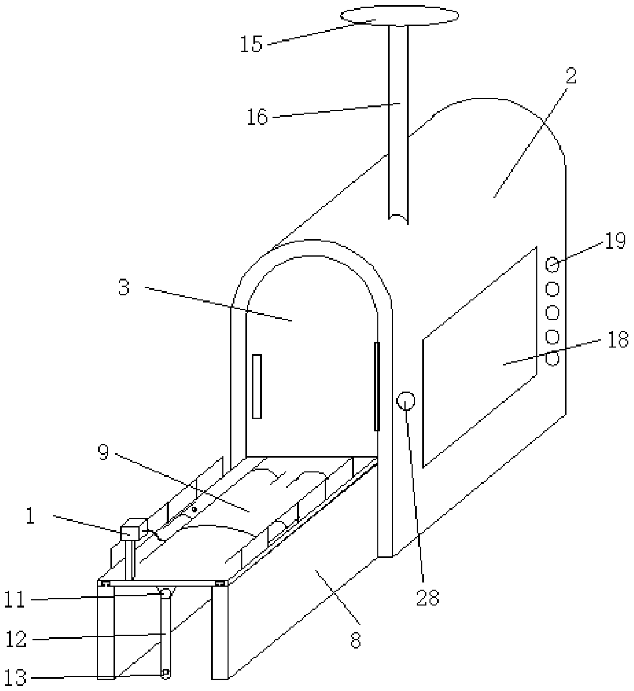
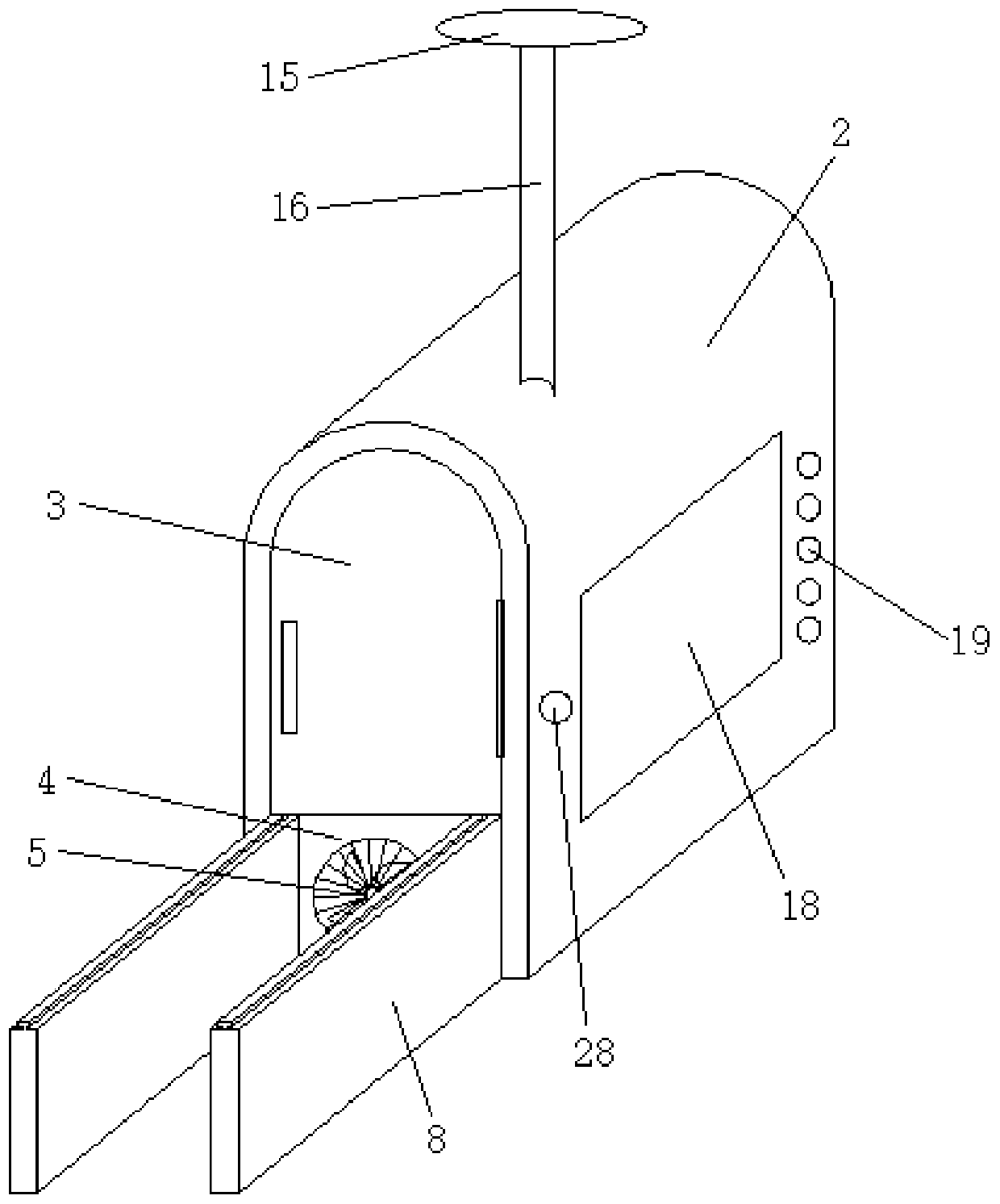
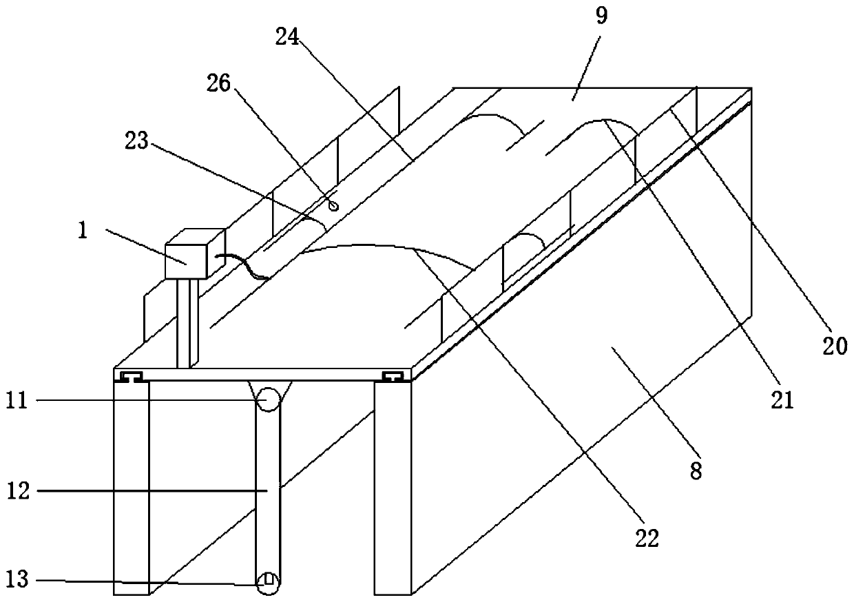
[0029] The present invention provides a cardiac angiography operation device which can automatically adjust the position for the Department of Cardiology, specifically as Figure 1 to Figure 5 As shown, it includes a contrast protection device, an external support device, a contrast bed, a contrast device, a contrast medium storage and injection device 1 and a monitoring device; in this embodiment, the contrast medium storage and injection device 1 is a contrast medium injection used in the existing cardiac contrast surgery Device, this part is not improved in this embodiment, and the existing contrast agent injection device can meet the requirements of this application.

[0030] The radiography protection device includes a metal shell 2, the metal material has the function of preventing X-rays, and a camera is arranged on the inner top of the metal shell 2, and the camera is used for real-time monitoring of the whole operation. The front side of the metal shell 2 is provided ...


PUM


Abstract

The invention provides a cardiography device capable of automatically adjusting the position for the cardiology department, and belongs to the technical field of medical tools. The cardiography devicecomprises a radiography protection device, an external supporting device, a radiography bed, a radiography device, a contrast medium storing and injecting device and a monitoring device. The radiography bed can be supported outside the radiography protection device through the external supporting device so that a preoperative preparation can be made for a patient, the radiography protection device is used for sealing the whole radiography bed for real-time radiography surgery and preventing X-rays from hurting medical staff, the adjustment of tilt angles of the two sides of the radiography bed is realized in the radiography protection device push rod motors, the radiography requirements at different angles and positions are met, a robot arm can rotate by 360 degrees and stretch out and draw back, the radiography device conducts precise radiography on the part, needing radiography, of the body of the patient through the robot arm, and the monitoring device is used for controlling and monitoring the whole radiography process. The cardiography device is easy and convenient to operate, saves time and labor, is scientific and effective and greatly relieves the work difficulty of the medical staff.

Description

technical field [0001] The invention belongs to the technical field of medical appliances, and in particular relates to a cardiac angiography operation device used in the department of cardiology and whose position can be automatically adjusted. Background technique [0002] Cardiovascular angiography is to insert a very thin catheter into the aorta of the human body, inject a contrast agent into the aortic blood through the small opening in front of the catheter, and inject the transparent contrast agent into the blood flow quickly under X-ray irradiation, so that the heart and The large blood vessel cavity is developed under X-ray irradiation. At the same time, there are methods such as rapid filming, TV photography, or tape video to record the development process of the heart and the large blood vessel cavity. From the results of the development, the blood flow sequence containing the contrast agent can be seen , and the filling of the great blood vessels of the heart, so...

Claims


Application Information

Patent Timeline
IPC IPC(8): A61B6/04A61B6/00A61B6/10
Inventor 付新司元元
Owner THE FIRST AFFILIATED HOSPITAL OF ZHENGZHOU UNIV
Learn More