
Patsnap Eureka
For R&D, Patsnap Eureka makes reading and utilizing patents & technical documents easy.
Patsnap Eureka AIR
Designed for self-driven R&D workflows. Generate viable solutions, solve complex R&D challenges, empower your innovation with AI.

Patsnap Eureka Materials
Designed for material experts only. Revolutionize your material R&D, from search, analyze, to developing new materials.

TechResearch
Generate reliable direction feasibility study reports for your R&D in just a few steps.

TechSeek
Discover and master advanced knowledge NOW. Basics, ideas, possibilities, all at once.

TechMind
As an expert in R&D Theories, TechMind can generates customized viable solutions instantly.

TechRisk
Analyze your overall solution with one click, know your potential R&D risks in advance.

TechMonitor
Get weekly tech updates, stay abreast of the latest tech innovations and key insights.
A pleural effusion discrimination method and device and a computer device
A discrimination method and technology of a discriminating device are applied in the field of image processing and can solve the problems of low efficiency, lack of efficiency and accuracy of pleural effusion discrimination method and the like.
- Summary
- Abstract
- Description
- Claims
- Application Information
AI Technical Summary
Problems solved by technology
Method used
Image

Examples
Embodiment 1
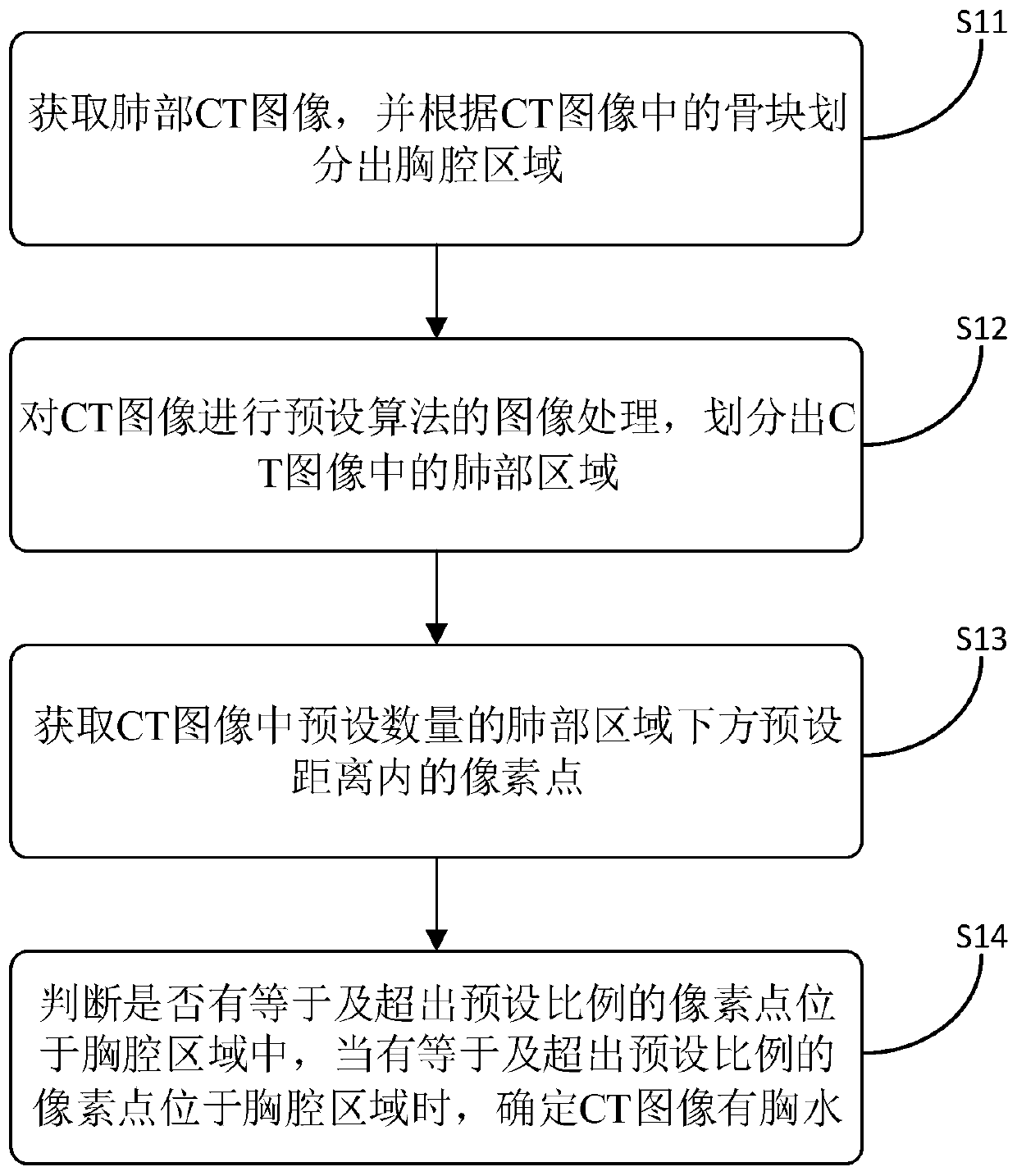
[0057] figure 1It is a flow chart of a method for discriminating pleural effusion provided in Embodiment 1 of the present invention, and the method includes the following steps:
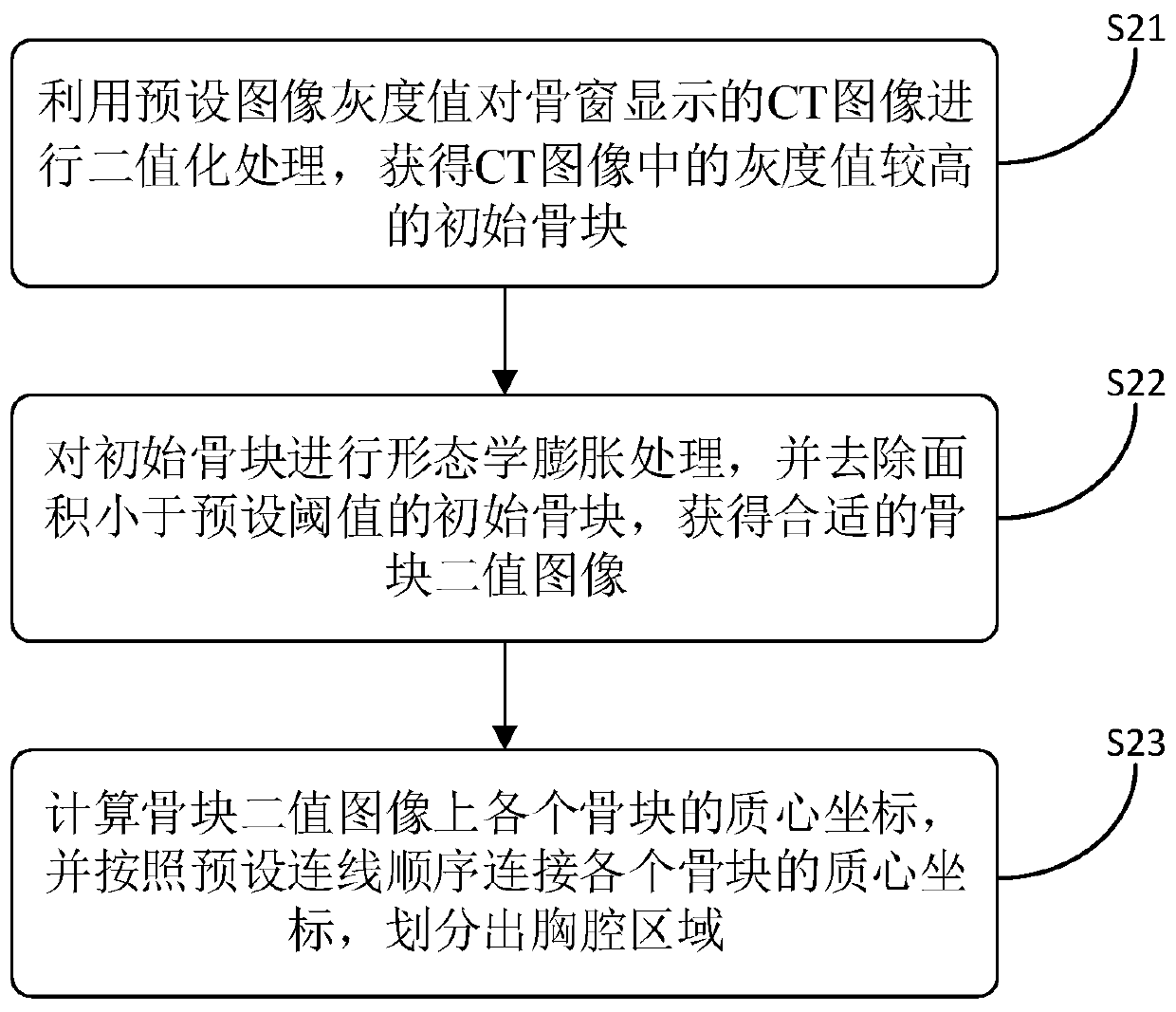
[0058] Step S11: Obtain a CT image of the lungs, and divide the chest region according to the bone fragments in the CT image.
[0059] In the embodiment of the present invention, the CT image (CT, Computed Tomography) of the lungs is mainly used to judge the pleural effusion, wherein the CT image is an electronic computerized tomography image, using precisely collimated X-ray beams, Y-rays and ultrasound, etc., Together with the extremely high-sensitivity detector, one by one cross-sectional scanning is performed around a certain part of the human body. Specifically, this embodiment is a cross-sectional scanning image of the human lung.
[0060] In the embodiment of the present invention, the lung CT images are obtained, and before the lung CT images are divided into regions, all lung CT images can ...
Embodiment 2
[0087] Figure 4 It is a flow chart of a method for discriminating pleural effusion provided in Embodiment 2 of the present invention, and the method includes the following steps:
[0088] Step S41: Obtain a CT image of the lungs, and divide the chest region according to the bone fragments in the CT image.
[0089] This step is consistent with the above step S11, and will not be repeated here.
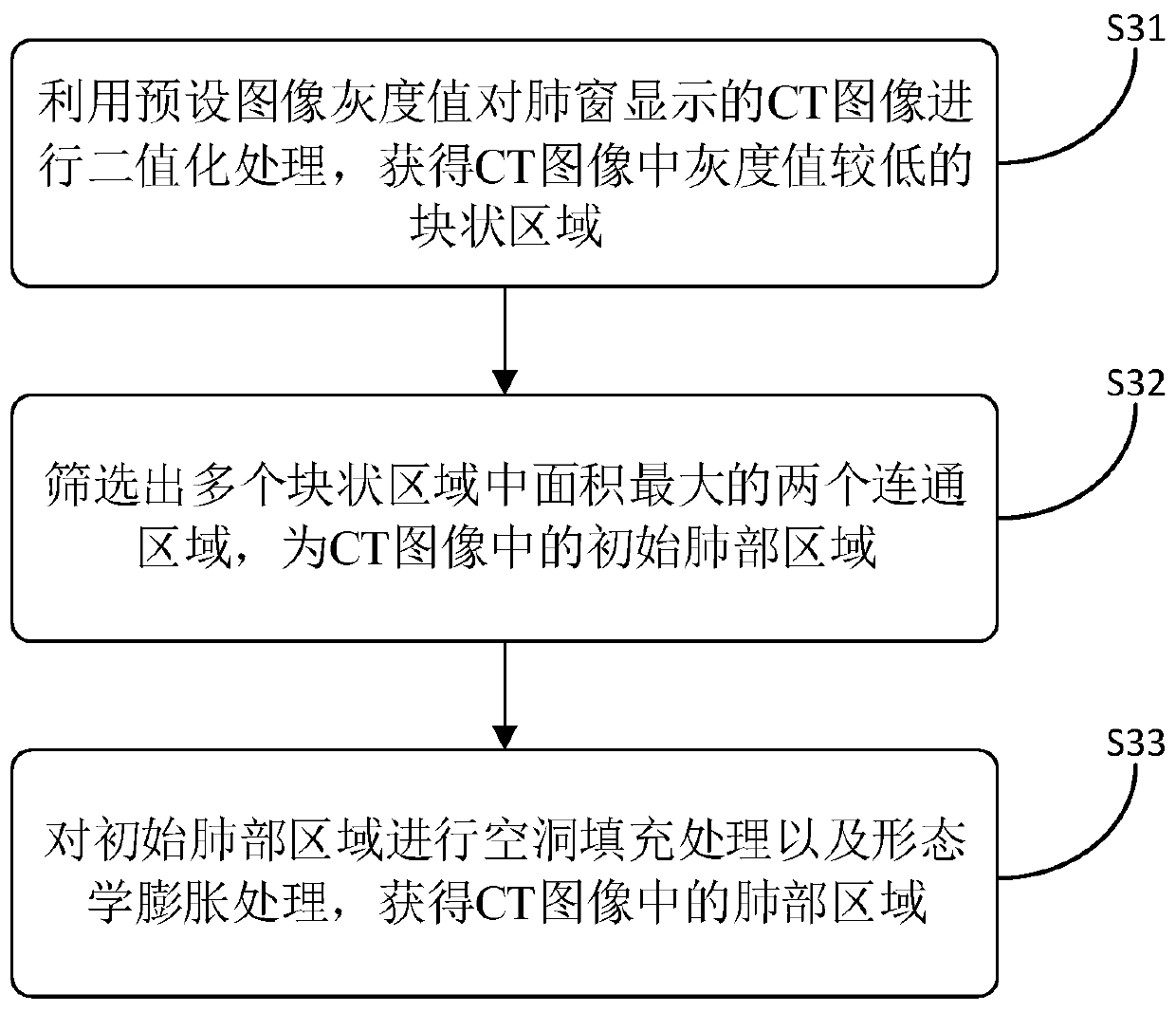
[0090] Step S42: Carry out image processing with a preset algorithm on the CT image, and divide the lung area in the CT image.
[0091] This step is consistent with the above step S12, and will not be repeated here.
[0092] Step S43: acquiring pixels within a preset distance below a preset number of lung regions in the CT image.
[0093] This step is consistent with the above step S13, and will not be repeated here.
[0094] Step S44: Determine whether there are pixel points equal to or exceeding the preset ratio located in the thoracic region, and when there are pixel points equal ...
Embodiment 3
[0106] Image 6 It is a schematic structural diagram of a pleural effusion discrimination device provided in Embodiment 3 of the present invention.
[0107] The pleural effusion discrimination device 600 includes:
[0108] The thorax region division module 610 is configured to acquire a lung CT image, and divide the thoracic region according to the bone blocks in the CT image.
[0109] The lung region division module 620 is configured to perform image processing of a preset algorithm on the CT image, and divide the lung region in the CT image.
[0110] A pixel point acquisition module 630, configured to acquire a preset number of pixel points within a preset distance below the lung area in the CT image.
[0111] The pleural effusion judging module 640 is used to judge whether there are pixel points equal to or exceeding the preset ratio located in the chest region, and when there are pixel points equal to or exceeding the preset ratio located in the chest region, it is deter...
PUM
 Abstract
Description
Claims
Application Information
 - R&D Engineer
- R&D Manager
- IP Professional
- Industry Leading Data Capabilities
- Powerful AI technology
- Patent DNA Extraction
Browse by: Latest US Patents, China's latest patents, Technical Efficacy Thesaurus, Application Domain, Technology Topic, Popular Technical Reports.
© 2024 PatSnap. All rights reserved.Legal|Privacy policy|Modern Slavery Act Transparency Statement|Sitemap|About US| Contact US: help@patsnap.com