Method for producing endothelial cells

A technology of endothelial cells and endothelial progenitor cells, applied in the direction of vascular endothelial cells, germ cells, biochemical equipment and methods, etc.

Pending Publication Date: 2019-07-09
TAKARA HOLDINGS
View PDF8 Cites 1 Cited by
  • Summary
  • Abstract
  • Description
  • Claims
  • Application Information

AI Technical Summary

Problems solved by technology

[0012] The present invention aims to solve the problems that conventional production methods have, and

Method used


Image

  • Method for producing endothelial cells
  • Method for producing endothelial cells
  • Method for producing endothelial cells

Examples


Embodiment 1

[0121] Example 1: Differentiation of iPS cells into vascular endothelial cells

[0122] Human iPS cells (ChiPSC12 strain) (Cellartis) were cultured in DEF-CS medium (Cellartis) according to the instructions attached to the medium.

[0123] In order to effectively induce the differentiation of mesoderm cells, iPS cells were coated with Matrigel according to the following procedure (Matrigel sandwich method). First, TrypLE (trademark) Select (Life technologies) was added to subcultured iPS cells, and the cells were incubated at 37°C for 3-7 minutes. Collect the separated cells as single cells by pipetting, and count the number of cells. Next, divide the cells into 6 x 10 4 Cells / cm 2 The density was inoculated into a culture vessel coated with Matrigel (trademark) (Corning) and cultured in DEF-CS medium for 2-3 days. After that, the medium was replaced with DEF-CS medium supplemented with Matrigel diluted to 1:60, and the cells were cultured for a further 16-24 hours to coat the up...

Embodiment 2

[0134] Example 2: Freezing and thawing of vascular endothelial cells

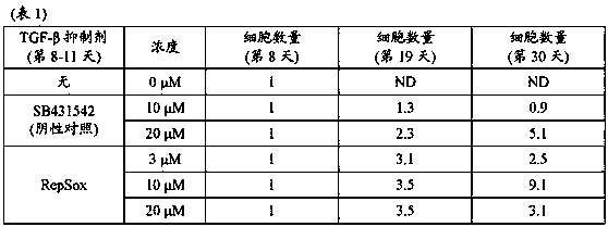
[0135] The induction of vascular endothelial cells was performed according to the method described in Example 1 using the medium supplemented with 20 μM RepSox, and the cells were collected from day 18-22. Wash the collected cells and suspend them in Stem Cell Banker (TAKARABIO INC.) so that the cell concentration is 3 x 10 6 / mL, and dispense 1 mL of each suspension in vials. The vial was placed in a refrigerated container (BICELL, Nihon Freezer Co., Ltd.), and the cells were subjected to slow freezing in a freezer storage chamber maintained at a temperature of -80°C. After that, each vial was transferred to liquid nitrogen and stored for 3 days.

[0136] The frozen cells were warmed in a water bath warmed to 37°C for 2 minutes and 30 seconds to thaw the cells. The cell viability was measured by using a part of the cells, and the result was that the cell viability was 87%. The entire amount of the remaining ...

Embodiment 3

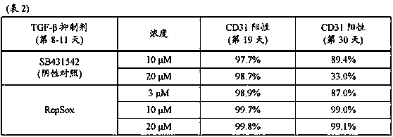
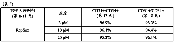
[0138] Example 3: Measurement of CD31+ / CD34+ cells

[0139] According to the instructions attached to the medium, human iPS cells (ChiPSC12 strain) were cultured in DEF-CS medium.

[0140] TrypLE (trademark) Select was added to iPS cells that had been subcultured, and the cells were incubated at 37°C for 3-7 minutes. Collect the separated cells as single cells by pipetting, and count the number of cells. Next, divide the cells into 6x 10 4 Cells / cm 2 The density was inoculated into a culture vessel coated with Matrigel (trademark), and cultured in DEF-CS medium for 2-3 days. After that, the medium was replaced with DEF-CS medium supplemented with Matrigel diluted to 1:60, and the cells were cultured for a further 16-24 hours to coat the upper layer of the cells with Matrigel.

[0141] The obtained Matrigel-coated iPS cells were differentiated into mesoderm cells according to the following procedure. First, the medium of Matrigel-coated iPS cells was replaced with RPMI 1640 medium ...


PUM


Abstract

The present invention relates to a method for producing endothelial cells, in which a step (a) of inducing a mesoblastic cell mass containing endothelial progenitor cells from pluripotent stem cells without forming embryoid bodies and a step (b) of culturing the mesoblastic cell mass containing the endothelial progenitor cells in the presence of RepSox are carried out in this order. According to the present invention, it becomes possible to produce high-quality endothelial cells from pluripotent stem cells with high efficiency. The endothelial cells produced by the method of the present invention are useful, for example, for the production of a myocardial cell sheet, and is expected to be used for the treatment of heart diseases. A myocardial cell sheet can be produced by mixing endothelial cells produced by the method of the present invention with myocardial cells and wall cells and then culturing the mixture.

Description

Technical field [0001] The present invention relates to a method for producing high-quality endothelial cells from pluripotent stem cells. Background technique [0002] Technological innovations related to regenerative therapy have been developed, especially methods for generating pluripotent stem cells and methods for inducing differentiation of pluripotent stem cells, so that research on regeneration of biological tissues has been intensively conducted. And for the heart, which is essential for the life maintenance of a single organism, it is believed that regenerative medicine technology can be used to treat heart diseases. For example, attempts have been made to apply cardiomyocytes artificially produced from pluripotent stem cells to the treatment of heart diseases. At this time, cardiomyocytes are not used alone, but, for example, a method for forming a laminar cell structure containing cardiomyocytes has been developed (Patent Publication 1), and the thus obtained cardiom...

Claims


Application Information

Patent Timeline
IPC IPC(8): C12N5/0735C12N5/071C12N5/10
CPCC12N5/069C12N2506/45C12N2533/90C12N2501/16C12N2501/165C12N2501/155C12N2501/115C12N2501/15A61P9/00C12N5/0602C12N2502/1329C12N2502/23C12N2506/02A01N1/0284C12N5/0692C12N2501/999
Inventor 山本由姬榎龙嗣户坂泰弘山口庸子峰野纯一
Owner TAKARA HOLDINGS
Why Eureka
Try Eureka