
Kidney medical image contour line extraction system and method

A medical image and extraction system technology, applied in the field of medical image extraction system, can solve the problems of slow calculation speed and complex extraction method of organ tissue outline, and achieve the effect of easy edge detection, fast calculation and simple method

Pending Publication Date: 2019-07-23
THE AFFILIATED HOSPITAL OF QINGDAO UNIV
View PDF2 Cites 1 Cited by
  • Summary
  • Abstract
  • Description
  • Claims
  • Application Information

AI Technical Summary

Problems solved by technology

However, the method for extracting the outline of organs and tissues based on medical images disclosed in this patent application is relatively complicated and the calculation speed is relatively slow.

Method used


Image

  • Kidney medical image contour line extraction system and method
  • Kidney medical image contour line extraction system and method
  • Kidney medical image contour line extraction system and method

Examples


Embodiment Construction

[0032] Embodiments of the present invention are described in detail below, examples of which are shown in the drawings, wherein the same or similar reference numerals designate the same or similar elements or elements having the same or similar functions throughout. The embodiments described below by referring to the figures are exemplary and are intended to explain the present invention and should not be construed as limiting the present invention.

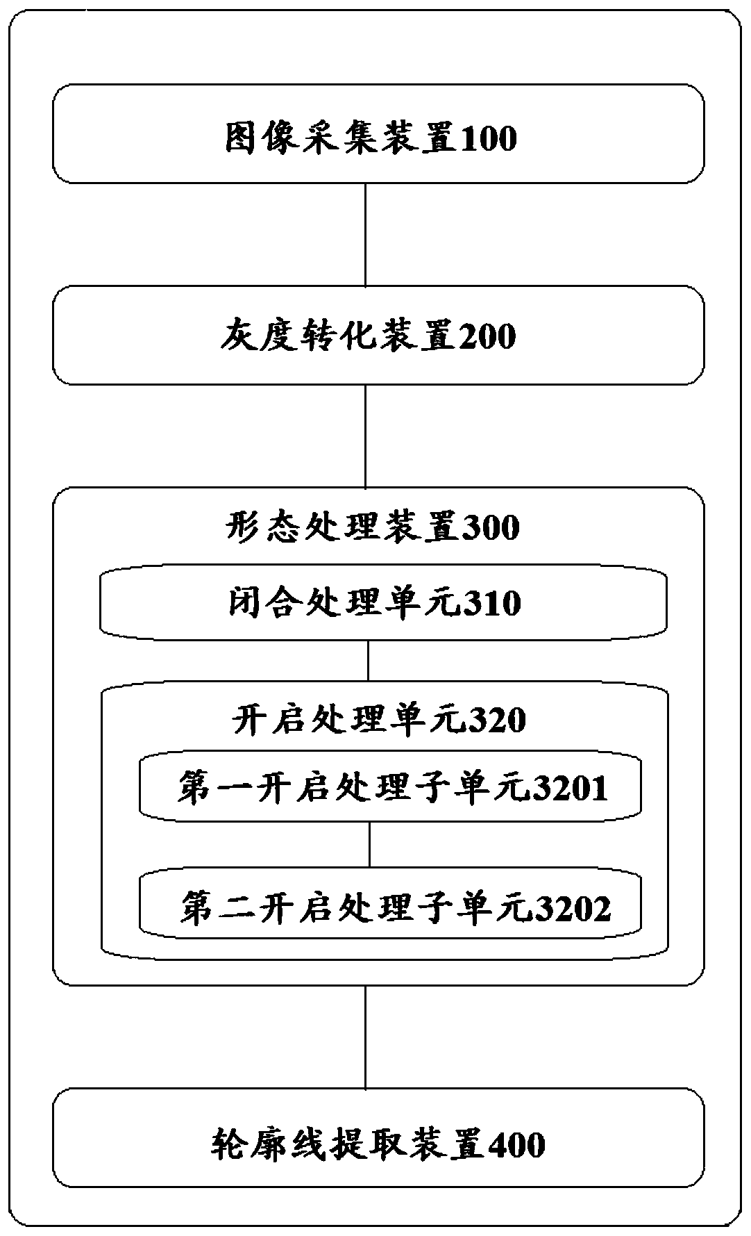
[0033] First, please refer to figure 1 , as a non-limiting implementation, the renal medical image contour extraction system of the present invention includes: an image acquisition device 100 , a grayscale conversion device 200 , a morphology processing device 300 and a contour line extraction device 400 .

[0034] Wherein, the image acquisition device 100 can obtain a CT image of a specific kidney, where the CT image may be a CT image of the specific kidney in a plain scan phase, an arterial phase, a venous phase, and a renal pe...


PUM


Abstract

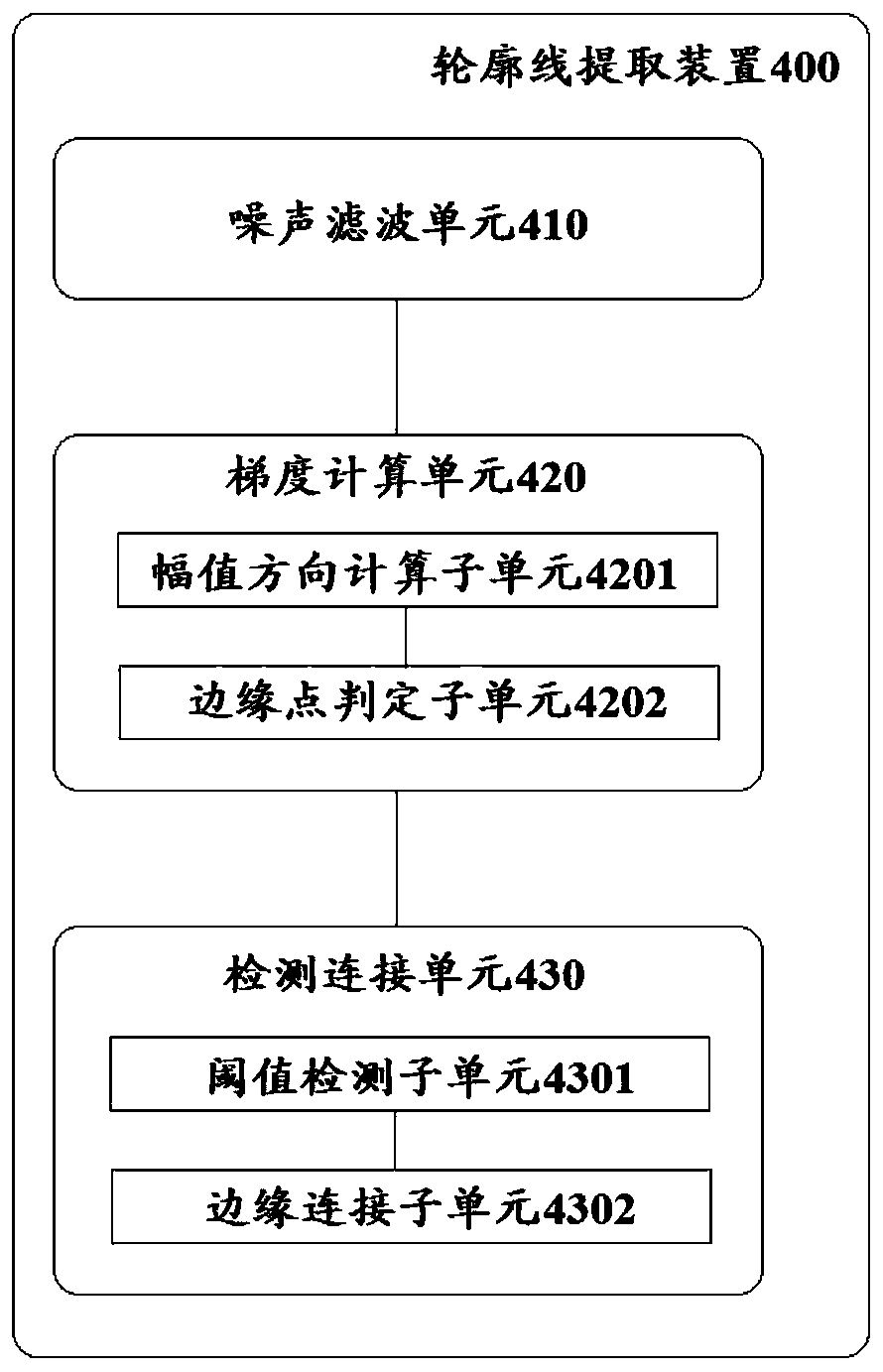
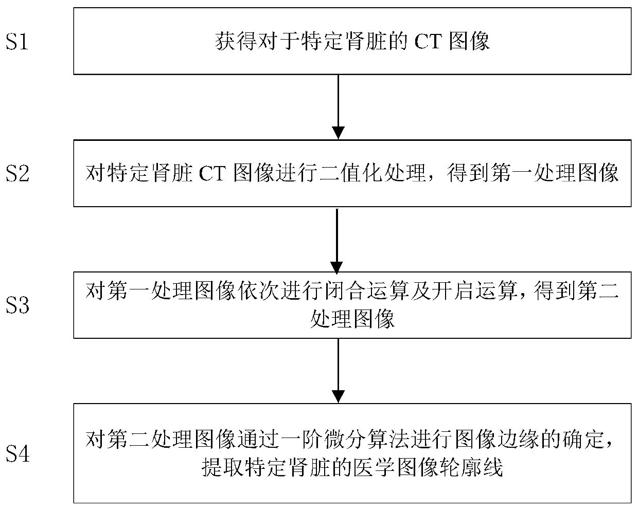
The invention discloses a kidney medical image contour line extraction system and method, and the system comprises an image collection device, a gray level conversion device, a shape processing device, and a contour line extraction device, which are in communication connection in sequence, and the image collection device is used for obtaining a CT image of a specific kidney; the gray level conversion device is used for carrying out binarization processing on the specific kidney CT image acquired by the image acquisition device to obtain a first processed image; the morphological processing device is used for sequentially carrying out closing operation and opening operation on the first processed image to obtain a second processed image; and the contour line extraction device is used for determining the image edge of the second processed image through a first-order differential algorithm so as to extract a medical image contour line of a specific kidney.

Description

technical field [0001] The invention relates to the technical field of medical image processing, in particular to a medical image extraction system. Background technique [0002] In recent years, renal CT images have become more and more widely used clinically, and have become one of the important means for doctors to conduct renal function, pathology and anatomy, and the determination of renal boundaries is a prerequisite for quantitative analysis. The automatic segmentation of renal CT sequences and the extraction of renal borders are of great help to reduce the workload of doctors and improve the speed and accuracy of diagnosis. [0003] For example, Chinese Patent Application Publication No. CN106108932A discloses a device and method for fully automatic kidney region-of-interest extraction, which initially divides the preliminary region of interest based on the level set C-V model algorithm, and extracts the suspected region of interest; The kidney body ratio parameters...

Claims


Application Information

Patent Timeline
Patent Type & Authority Applications(China)
IPC IPC(8): G06T7/00G06T7/13G06T7/136G06T7/149
CPCG06T7/0012G06T7/13G06T7/136G06T7/149G06T2207/10081G06T2207/30084
Inventor 魏宾董蒨朱呈瞻董冰子夏楠王菲菲王凤娇楚宏硕
Owner THE AFFILIATED HOSPITAL OF QINGDAO UNIV
Learn More