AR and endoscope combined surgical navigation method and terminal

A surgical navigation and endoscope technology, applied in the medical field, can solve problems such as the inability to intuitively understand the surrounding conditions of the endoscope

Inactive Publication Date: 2019-07-26
厦门强本科技有限公司
View PDF6 Cites 8 Cited by
  • Summary
  • Abstract
  • Description
  • Claims
  • Application Information

AI Technical Summary

Problems solved by technology

[0003] The endoscopes currently on the market are basically displayed in two-dimensional pictu

Method used


Image

  • AR and endoscope combined surgical navigation method and terminal
  • AR and endoscope combined surgical navigation method and terminal
  • AR and endoscope combined surgical navigation method and terminal

Examples


Embodiment 1

[0066] Please refer to Figure 1 to Figure 3 , Embodiment 1 of the present invention is:

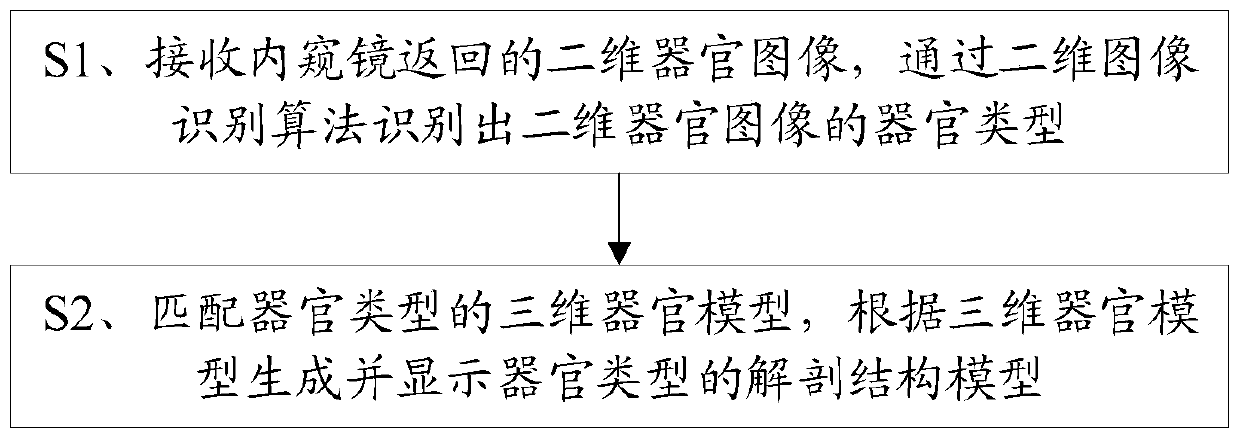
[0067] The surgical navigation method combining AR and endoscope, including steps:

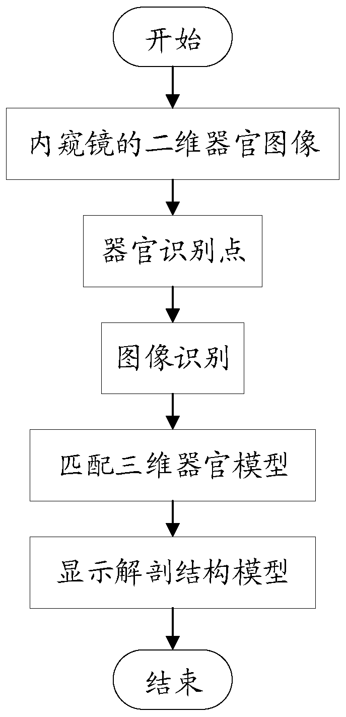
[0068] S1. Receive the two-dimensional organ image returned by the endoscope, and identify the organ type of the two-dimensional organ image through a two-dimensional image recognition algorithm;

[0069] Wherein, after receiving the two-dimensional organ image returned by the endoscope in step S1, it also includes: finding the organ identification point corresponding to the organ feature from the two-dimensional organ image;

[0070] S2. Obtain the three-dimensional organ model of the organ type, generate and display the anatomical structure model of the organ type according to the three-dimensional organ model;

[0071] Wherein, in step S2, the anatomical structure model generated and displayed according to the three-dimensional organ model is specifically:

[0072] The position and direction of the...

Embodiment 2

[0079] Please refer to Figure 1 to Figure 3 , the second embodiment of the present invention is:

[0080] The AR and endoscope combined surgical navigation method, on the basis of the first embodiment above, further includes steps after step S2:

[0081] S3. Simultaneously display the two-dimensional organ image and the anatomical structure model, and display the position and direction of the endoscope in the organ on the interface where the anatomical structure model is located;

[0082] S4. Obtain operation instructions, and operate the anatomical structure model according to the operation instructions. The operations include movement, overall scaling, transparent control, and one-key restoration.

[0083] In this embodiment, the virtual organ can be operated through the keyboard, wherein, the Q key is to move the position of the virtual model; the S key is to zoom the virtual model as a whole; the A key is to transparently control the position of the virtual model; R The...

Embodiment 3

[0084] Please refer to Figure 4 as well as Figure 5 , Embodiment three of the present invention is:

[0085] The combined AR and endoscope surgical navigation terminal 1 includes a memory 3, a processor 2, and a computer program stored in the memory 3 and operable on the processor 2. When the processor 2 executes the computer program, it realizes the above-mentioned first embodiment. step.

[0086] Such as Figure 5 As mentioned above, the AR and endoscope combined surgical navigation terminal 1 preferably uses a Zspace display screen, and a set of software developed by unity is installed in the Zspace display screen to realize the steps in the first embodiment above. Configure the parameters on the Zspace display, connect the endoscope through the USB interface, open the software, and select the model of the endoscope to be viewed. At this time, the Zspace display can receive the secondary data seen on the endoscope Dimensional organ images can also be connected to a TV...


PUM


Abstract

The invention discloses an AR and endoscope combined surgical navigation method and terminal. The method includes receiving a two-dimensional organ image returned by an endoscope and identifying an organ type of the two-dimensional organ image by a two-dimensional image identification algorithm; acquiring a three-dimensional organ model of the organ type and generating and displaying an anatomicalmodel of the organ type according to the three-dimensional organ model. The three-dimensional virtual anatomical model is generated through a two-dimensional organ image. On one hand, a doctor can see a real image presented by the two-dimensional organ image; on the other hand, a virtual image formed by the anatomical model can be seen, that is, the virtual reality contrast technology is adopted,so that the doctor can have a more intuitive understanding of the surrounding conditions of the endoscope, and the effect of surgical navigation can be achieved.

Description

technical field [0001] The invention relates to the field of medical technology, in particular to a surgical navigation method and a terminal combining AR and endoscope. Background technique [0002] Endoscope is a detection instrument that integrates traditional optics, ergonomics, precision machinery, modern electronics, mathematics and software. It includes image sensors, optical lenses, light source lighting, mechanical devices, etc. It can enter the stomach through the mouth or enter the body through other natural channels, or it can enter the human body through a small incision made through surgery. In modern medicine, endoscopes can be used to see lesions that cannot be shown by X-rays. For example, doctors can observe ulcers or tumors in the stomach with the help of endoscopes, and formulate the best treatment plan accordingly. [0003] The endoscopes currently on the market are basically displayed in two-dimensional pictures, and it is impossible to intuitively und...

Claims


Application Information

Patent Timeline
IPC IPC(8): A61B34/20A61B34/10G06T19/00
CPCA61B34/10A61B34/20A61B2034/105A61B2034/2065G06T19/006
Inventor 王博亮
Owner 厦门强本科技有限公司
Why Eureka
Try Eureka