MRI/CT fusion method based on bending wave

A fusion method and bending wave technology, applied in image enhancement, image analysis, instruments, etc., can solve the problems of ineffective information fusion and inability to extract medical image structure information well, so as to achieve good capture of image curves and short time , good effect

Active Publication Date: 2020-12-29
ZHEJIANG HOSPITAL
View PDF8 Cites 0 Cited by
  • Summary
  • Abstract
  • Description
  • Claims
  • Application Information

AI Technical Summary

Problems solved by technology

[0008] In the prior art, many classic multi-scale transformation methods have played an important role in image fusion, but these fusion methods used in ordinary natural images often cannot extract useful structural information of medical images, which leads to medical image fusion. The useful information cannot be well integrated into the final generated image during image fusion

Method used


Image

  • MRI/CT fusion method based on bending wave
  • MRI/CT fusion method based on bending wave
  • MRI/CT fusion method based on bending wave

Examples


Embodiment Construction

[0059] The present invention will be further described below in conjunction with the accompanying drawings.

[0060] The fusion algorithm of medical MRI / CT images based on bending wave transform (bentlet transform), the steps are as follows:

[0061] Step 1) Construct a PET / CT image model.

[0062] We assume that the PET / CT image is composed of the real part of the image and the multiplicative noise of the image. Usually, in order to compress the image signal, the generated medical image will be logarithmically transformed, and the original multiplicative noise will become additive noise. The final MRI image The model is as follows:

[0063] m(u,v)=i(u,v)+z(u,v) (1)

[0064] The model of the CT image is as follows:

[0065] c(u,v)=i(u,v)+z(u,v) (2)

[0066] Where (u, v) represents the coordinate values ​​of the MRI image and CT image, i(u, v) represents the real signal, and z(u, v) represents the additive noise.

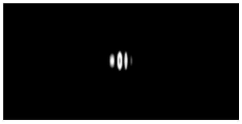
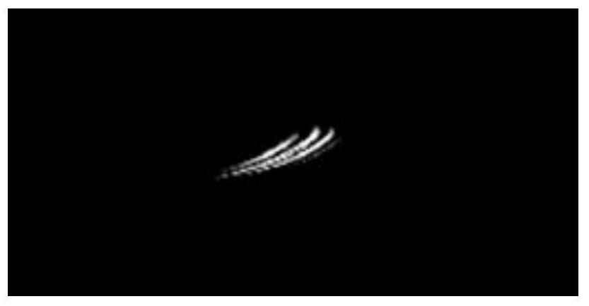
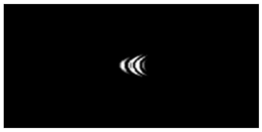
[0067] Step 2) Construct the bending wave system.

[0068...


PUM


Abstract

The invention discloses a medical MRI / CT image fusion method based on bending wave transformation. The method comprises the following steps: 1) constructing a PET / CT image model; 2) constructing a bending wave system; 3) performing bending wave transformation on the MRI image and the CT image; 4) fusing the MRI image bending wave coefficient obtained in the step 3) and the CT image bending wave coefficient; and step 5) carrying out bending wave inverse transformation on a bending wave coefficient obtained after the bending wave coefficients of the two images are fused. According to the method,experimental analysis is compared with an image fusion method based on common weighted average, and the method is effectively applied to the field of fusion of medical MRI / CT images; curved wave transformation is a novel transformation domain method, theoretical support of the method is developed by second-order shear wave transformation, one more bending variable exists in function parameters ofthe method, and curve information in a captured image can be better fitted through a curved wave basis function. Through comparison of experimental data, a medical MRI / CT image fusion algorithm basedon bending wave transformation is provided, and analysis and diagnosis of doctors can be better facilitated.

Description

technical field [0001] The invention relates to a medical MRI / CT fusion method. Background technique [0002] With the development of science and technology, in the field of medical imaging, imaging technologies such as ultrasound imaging, CT, and MRI have been applied in medical clinical diagnosis. Because MRI and CT have two different imaging principles combined in the same machine, it is not a simple addition of their functions. Instead, image fusion is performed on this basis. The fused image has both fine anatomical structure and rich physiological and biochemical function information, which can provide a basis for quantitative and qualitative diagnosis of the precise location of tumors and other lesions. X-rays can be used to perform attenuation correction on nuclear medicine images. [0003] Computed Tomography (Computed Tomography) uses precisely collimated X-ray beams, γ-rays, ultrasonic waves, etc., together with highly sensitive detectors, to conduct cross-secti...

Claims


Application Information

Patent Timeline
Patent Type & Authority Patents(China)
IPC IPC(8): G06T5/50G06T5/00
CPCG06T5/002G06T5/50G06T2207/10081G06T2207/10088G06T2207/20064G06T2207/30004
Inventor 程芸吕金城周海林陈坚张聚
Owner ZHEJIANG HOSPITAL
Why Eureka
Try Eureka