
A method for detecting developmental toxicity of chemicals

A technology of toxicity detection and detection method, applied in the direction of measuring devices, scientific instruments, biological testing, etc., can solve the problems of research, many influencing factors, and long experiment cycle

Active Publication Date: 2021-07-30
SHANGHAI NINTH PEOPLES HOSPITAL SHANGHAI JIAO TONG UNIV SCHOOL OF MEDICINE
View PDF9 Cites 0 Cited by
  • Summary
  • Abstract
  • Description
  • Claims
  • Application Information

AI Technical Summary

Problems solved by technology

However, there are several problems that cannot be ignored in carrying out animal experiments: 1. It requires a lot of financial resources; 2. The experiment cycle is long; 3. It requires a large number of experimental animals; 4. It is difficult to carry out metabolism and mechanism research due to many influencing factors in the body
[0005] Constrained by medical ethics, at present, the international safety evaluation testing methods are still based on animals and cells. Except for newly developed drugs whose safety is guaranteed by the results of pre-clinical experiments and have great therapeutic potential, almost No Chemical Substances Allowed for "Human Trials"

Method used


Image

  • A method for detecting developmental toxicity of chemicals
  • A method for detecting developmental toxicity of chemicals
  • A method for detecting developmental toxicity of chemicals

Examples


Embodiment 1

[0100] The cultivation of embodiment 1 human embryonic stem cells

[0101] hES will be provided in a state of normal growth after recovery. When the cell density reaches more than 70%, the cells are inoculated into Matrigel pre-coated (self-prepared, 1:20 dilution) culture dishes in the form of clumps.

[0102] (1) Passage of hES: Cells were passaged using cell separation medium A.

[0103] On the 4th to 5th day after hES inoculation, the proliferation of stem cells reached a certain level, and colonies with larger diameters and higher densities (about 70%) could be observed in the culture dish, at which time they could be subcultured.

[0104] Aspirate the hES medium to be passaged and wash it twice with D-PBS.

[0105] Add 2ml of cell separation solution A to the culture dish, incubate in a 37°C, 5% CO2 incubator, take it out for about 20-25 minutes, it can be observed that the clone structure is obviously loose, and when the outer edge of the clone appears curled, add 6ml ...

Embodiment 2

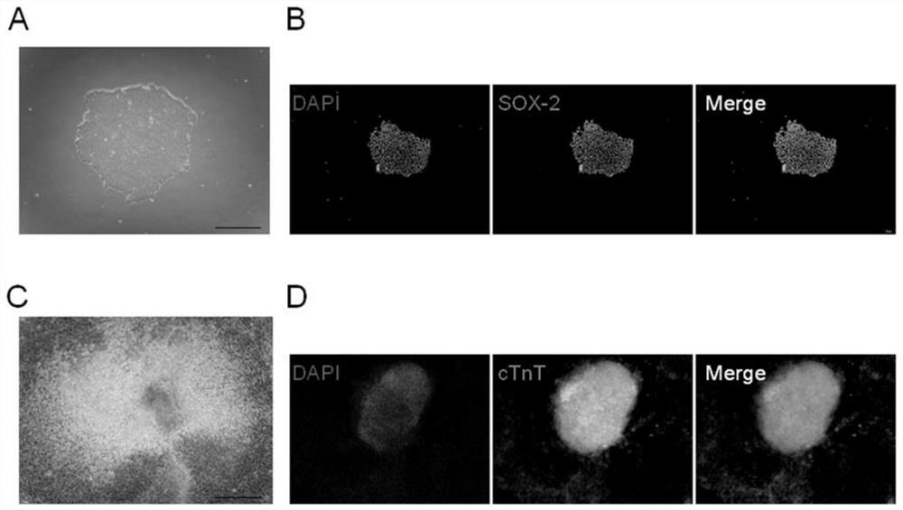
[0107] Example 2 Identification of Pluripotency of Human Embryonic Stem Cells

[0108] In this example, the identification of pluripotency of human embryonic stem cells was achieved by rapid alkaline phosphatase (AKP) staining (the kit needs to be purchased separately).

[0109] (1) Configure 5ml of 100mmol / L Tris HCl, adjust the pH to 8.2, add the dyeing reagent to it according to the kit instructions, and mix well for later use; absorb the culture medium in the ES cell culture dish, and use a high-temperature and high-pressure sterilized Wash with PBS, 5 minutes / time, 3 times in total, to remove the influence of the culture medium on the staining effect;

[0110] (3) Add an appropriate amount of staining reagent to the culture dish, and incubate in a 37°C incubator for 20 minutes in the dark;

[0111] (4) After washing with PBS, add 4% paraformaldehyde, wash with TBST after 2 minutes, and observe on the microscope.

Embodiment 3

[0112] Example 3 Induced differentiation of human embryonic stem cells into cardiomyocytes

[0113] When the cells passaged by cell separation medium A grow to 70%, use 10ml of culture medium #2 per 10cm dish, place at 37°C, 5% CO 2 Cells continued to grow in the incubator.

[0114] The day 1 of differentiation was recorded when the culture medium #2 was changed.

[0115] On day 3 of differentiation, continue to culture cells using 10 ml of medium #3 per 10 cm dish.

[0116] On the 6th day of differentiation, use 10ml of culture medium #4 for each 10cm dish to continue culturing the cells, and replace the culture medium once on the 9th day, 11th day, and 15th day of differentiation respectively.

[0117] Cells differentiated from day 15 to day 20 were used to test the developmental toxicity of compounds.


PUM


Abstract

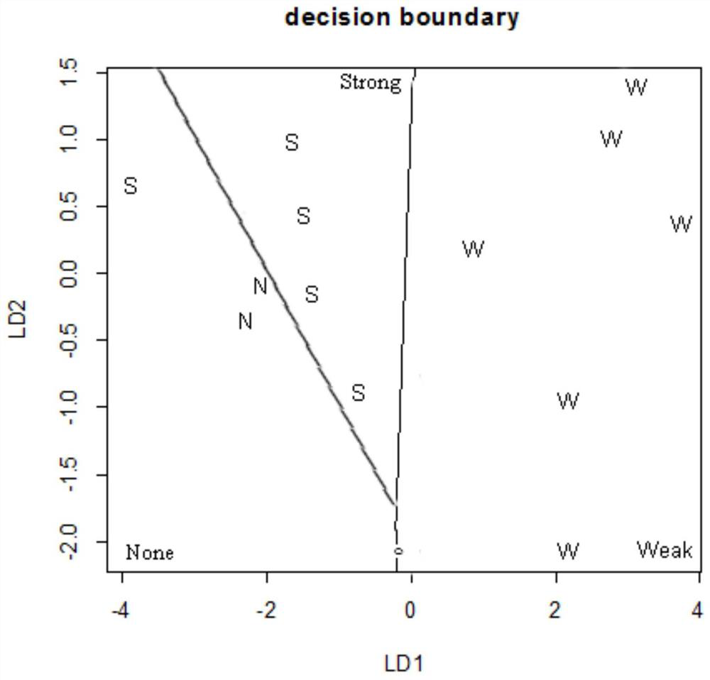
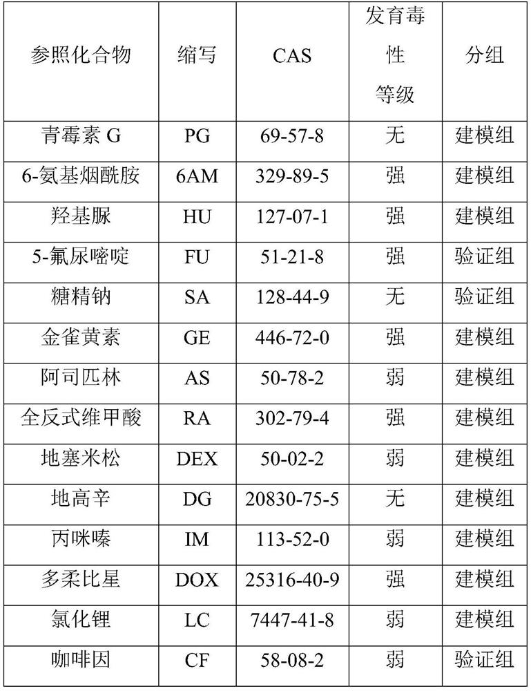
The invention provides a method for detecting the developmental toxicity of chemicals, which at least includes the following steps: respectively inducing undifferentiated human embryonic stem cells to differentiate into cardiomyocytes under the culture conditions of the chemical to be tested and without the chemical to be tested; There is the expression level of MYL4 in the cardiomyocytes under the culture conditions of the chemicals to be tested and without the chemicals to be tested; The change in the expression level of MYL4 in is ID; according to the value of ID, combined with the Fisher discriminant function equation, the developmental toxicity of the chemical to be tested can be judged. The invention uses human embryonic stem cells to establish a developmental toxicity test model for the first time, and the developmental toxicity information of the compound can be obtained through in vitro tests in a relatively short period of time; the invention can quickly detect and prompt whether a certain chemical has developmental toxicity information for humans.

Description

technical field [0001] The invention relates to the field of chemical toxicity detection, in particular to a chemical developmental toxicity detection method. Background technique [0002] At present, the toxicity data of chemicals are mostly obtained in experimental animals, such as acute toxicity test, subacute toxicity test, chronic toxicity test and so on. However, there are several problems that cannot be ignored in carrying out animal experiments: 1. It requires a lot of financial resources; 2. The experiment period is long; 3. It requires a large number of experimental animals; 4. It is difficult to carry out metabolism and mechanism research due to many influencing factors in the body. For the sake of animal protection, the "3R" principle is vigorously advocated, that is, reduction (Reduction), optimization (Refinement) and replacement (Replacement), whether it is from a scientific or economic point of view, toxicological alternatives are used to evaluate the hazards...

Claims


Application Information

Patent Timeline
Patent Type & Authority Patents(China)
IPC IPC(8): G01N33/50G01N33/533G01N33/58
CPCG01N33/5014G01N33/5061G01N33/533G01N33/582
Inventor 王艳程薇周韧杨守飞冯艳
Owner SHANGHAI NINTH PEOPLES HOSPITAL SHANGHAI JIAO TONG UNIV SCHOOL OF MEDICINE
Learn More