Method for pylorus and ileocecal valve positioning through wireless capsule endoscope images

A technology of capsule endoscopy and positioning method, applied in the field of image processing, to achieve the effect of promoting practical value, reducing work intensity, improving work efficiency and diagnosis rate

Active Publication Date: 2019-10-25
HANGZHOU DIANZI UNIV
View PDF20 Cites 11 Cited by
  • Summary
  • Abstract
  • Description
  • Claims
  • Application Information

AI Technical Summary

Problems solved by technology

[0003] However, wireless capsule endoscopy will generate a large amount of image da

Method used


Image

  • Method for pylorus and ileocecal valve positioning through wireless capsule endoscope images
  • Method for pylorus and ileocecal valve positioning through wireless capsule endoscope images
  • Method for pylorus and ileocecal valve positioning through wireless capsule endoscope images

Examples


Embodiment Construction

[0039] The present invention will be described in detail below in conjunction with the accompanying drawings and specific embodiments.

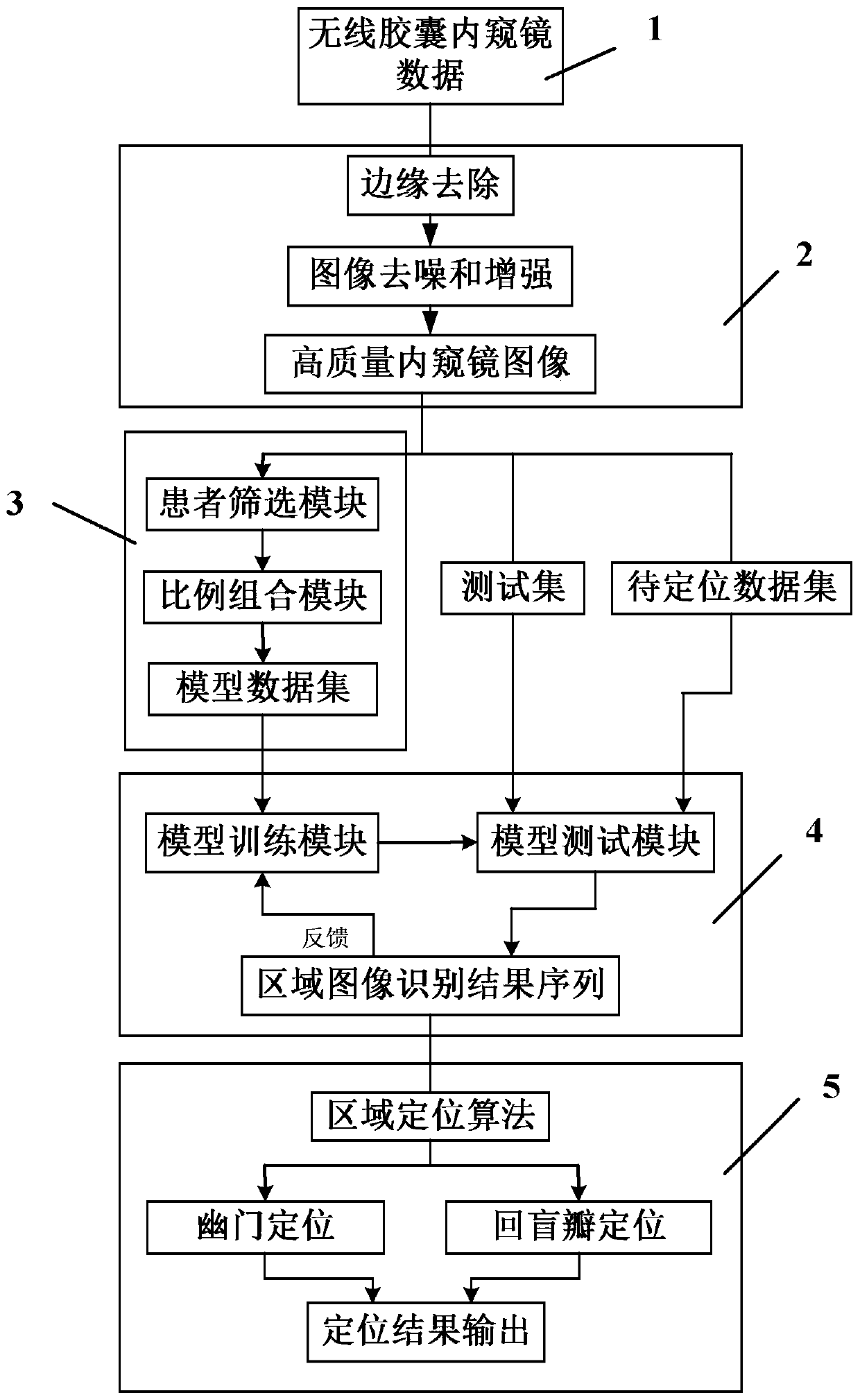
[0040] Such as figure 1 As shown, the wireless capsule endoscope image pylorus and ileocecal valve positioning method uses algorithm modules, including data input module 1, image preprocessing module 2, data sorting module 3, model adjustment module 4 and area positioning module 5; data The output of the input module 1 is connected to the input of the image preprocessing module 2, the output of the image preprocessing module 2 is connected to the input of the data sorting module 3, the output of the data sorting module 3 is connected to the input of the model adjustment module 4, and the model The output end of the adjustment module 4 is connected to the input end of the area positioning module 5 .

[0041] The data input module 1 obtains the wireless capsule endoscope video data of the patient, and obtains the wireless capsule endoscope ima...


PUM


Abstract

The invention discloses a method for pylorus and ileocecal valve positioning through wireless capsule endoscope images. When a wireless capsule endoscope is used for examination, the starting point and the stopping point of the small intestine are accurately found so that the workload of a doctor for viewing the images can be reduced, and missed diagnosis is reduced. According to the method, a deep learning thought serves as a technical core, a transfer learning strategy is also used, and a convolution neural network algorithm in a deep learning model is used for building an area image classifier; features of the wireless capsule endoscope images are obtained through automatic learning by means of training of the model, and then an area image identification result sequence is analyzed through an area positioning algorithm, so that pylorus and ileocecal valve positioning in the wireless capsule endoscope images is achieved. The method makes up for the blank of an existing capsule endoscope in the field of intelligent identification and accurate positioning, the working intensity of a doctor is greatly reduced, the working efficiency and the diagnosis rate are improved, the practicalvalue of the capsule endoscope in the clinical diagnosis of digestive tract diseases is further promoted, and a more efficient and more standard diagnosis mode is formed.

Description

technical field [0001] The invention belongs to the technical field of image processing, and in particular relates to a method for locating pylorus and ileocecal valve in a wireless capsule endoscope image. Background technique [0002] At present, conventional hand-held endoscopy in clinical application cannot involve the entire digestive tract. Gastroscopy can examine the upper digestive tract, while colonoscopy can only examine the colon and rectum, so there are still parts of the intestinal tract that conventional endoscopes cannot detect. and arrived. In recent years, with the development of science and technology, Wireless Capsule Endoscopy (WCE) has been widely used clinically, and has gradually become the mainstream technology for doctors to detect intestinal lesions. Usually, the patient swallows the capsule endoscope from the mouth, and through the peristalsis of the gastrointestinal tract, the capsule endoscope stays in the body for about 8 hours and takes images...

Claims


Application Information

Patent Timeline
IPC IPC(8): A61B1/04A61B1/273G06K9/62G06N3/04
CPCA61B1/041A61B1/273A61B1/00009A61B1/00016G06N3/045G06F18/24G06F18/214
Inventor 范姗慧刘士臣厉力华
Owner HANGZHOU DIANZI UNIV
Why Eureka
Try Eureka