Anti-PD-L1/anti-LAG3 bispecific antibody and uses thereof
A bispecific antibody, PD-L1 technology, applied in the direction of antibody, anti-receptor/cell surface antigen/cell surface determinant immunoglobulin, anti-animal/human immunoglobulin, etc., can solve the death of infected mice rate increase, etc.
- Summary
- Abstract
- Description
- Claims
- Application Information
AI Technical Summary
Problems solved by technology
Method used
Image
Examples
example
[0267] Hereinafter, the present invention will be described in detail by way of examples.
[0268] The following examples are only intended to illustrate the invention and should not be construed as limiting the invention.
example 1
[0269] Example 1: Preparation of anti-PD-L1 monoclonal antibody
[0270] 1.1. Preparation and analysis of anti-human PD-L1 mouse monoclonal antibody
[0271] Anti-human PD-L1 mouse monoclonal antibody was generated using hybridoma technology.
[0272] Antigen: human PD-L1-Fc protein and CHOK1 cell line with high expression of human PD-L1 (PDL1-CHOK1 cell line).
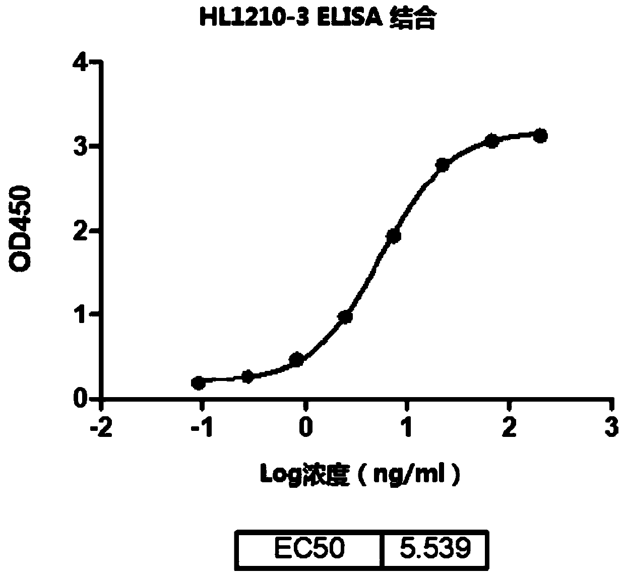
[0273] Immunization: To generate mouse monoclonal antibody against human PD-L1, first use 1.5x 10 7 6-8 week old female BALB / c mice were immunized with PDL1-CHOK1 cells. On day 14 and day 33 after the first immunization, use 1.5x 10 7 The immunized mice were re-immunized with PDL1-CHOK1 cells. To select mice that produced antibodies that bound the PD-L1 protein, sera from immunized mice were tested by ELISA. Briefly, microtiter plates were coated with 1 μg / ml human PD-L1 protein in PBS overnight at 100 μl / well at room temperature (RT), and then blocked with 100 μl / well of 5% BSA. Dilutions of plasma from immuniz...
example 2
[0376] Example 2. Preparation of anti-LAG3 monoclonal antibody
[0377] 2.1. Screening of fully human monoclonal antibodies against LAG-3
[0378] Anti-LAG3 human monoclonal antibody (α-LAG-3 mAb) was generated by screening a fully human Fab phage display library. The wild-type LAG-3-ECD-huFc fragment can bind to Daudi cells, while the D1-D2 truncated LAG-3-ECD-huFc fragment cannot bind to Daudi cells ( Figure 21 ). Therefore, the D1-D2 domain is critical for LAG-3 function.
[0379] Antigens for phage display library panning. LAG-3 is a single-pass type I membrane protein that belongs to the immunoglobulin (Ig) superfamily and contains four extracellular Ig-like domains (ECD): domains (D)1, D2, D3 and D4. Expression of recombinant human LAG-3-ECD-human IgG1 (LAG-3-huFc) fusion protein or human D1-D2 truncated LAG-3-ECD-human IgG1 (ΔD1D2-LAG-3-huFc) fusion protein in 293T cells system.
[0380] Phage library. Ig gene segments in mammals are arranged in groups of variab...
PUM
 Abstract
Description
Claims
Application Information
 - R&D
- Intellectual Property
- Life Sciences
- Materials
- Tech Scout
- Unparalleled Data Quality
- Higher Quality Content
- 60% Fewer Hallucinations
Browse by: Latest US Patents, China's latest patents, Technical Efficacy Thesaurus, Application Domain, Technology Topic, Popular Technical Reports.
© 2025 PatSnap. All rights reserved.Legal|Privacy policy|Modern Slavery Act Transparency Statement|Sitemap|About US| Contact US: help@patsnap.com


