Medical image processing device and method using convolutional neural network
A convolutional neural network and image data technology, applied in the field of medical image processing devices, can solve problems such as difficult technical solutions, inability to use nuclear magnetic resonance imaging, huge computational load, etc., and achieve the effect of improving diagnostic efficiency
- Summary
- Abstract
- Description
- Claims
- Application Information
AI Technical Summary
Problems solved by technology
Method used
Image
Examples
Embodiment approach 1
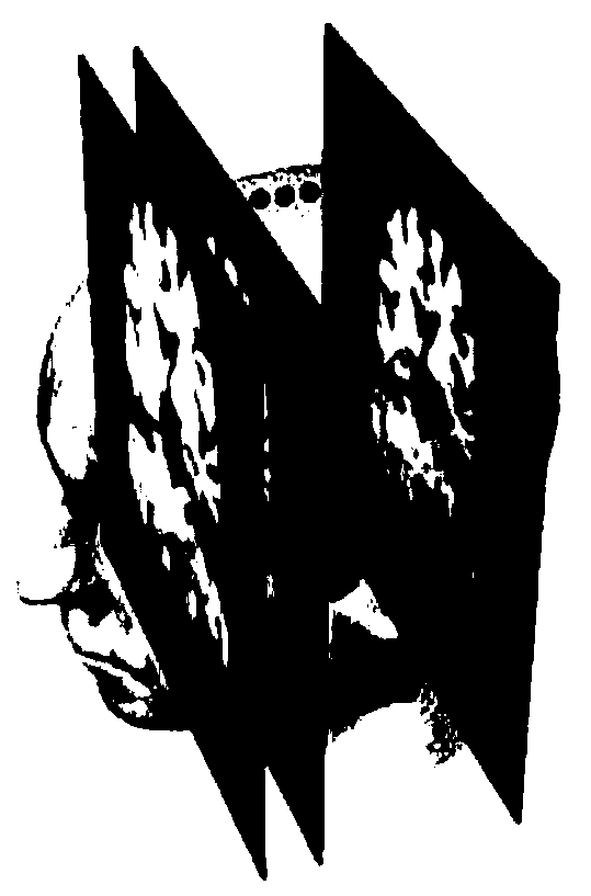
[0022] Embodiment 1 Utilizes the data of the Alzheimer's Disease Neuroimaging Initiative (ADNI for short) in the United States to realize the image processing and analysis of MRI in the present invention on the MRI image data of the cranium and the image data of the whole brain technical solutions. Download cranial MRIs of clinically confirmed Alzheimer's disease (AD), amnestic mild cognitive impairment (MCI) and normal aged group (NC) from the official website of the Alzheimer's Disease Neuroimaging Program in the United States image data,
[0023] Using the data of the Alzheimer's disease neuroimaging project in the United States, the 3T MRI image data of 450 subjects can be selected, and the training set, verification set and test set can be divided. The data in the training set is 313 subjects. 3T nuclear magnetic resonance image data, the data of the verification set is the 3T nuclear magnetic resonance image data of 120 subjects, and the 3T nuclear magnetic resonance im...
Embodiment approach 2
[0042] Embodiment 2 Utilizes the data of the Alzheimer's Disease Neuroimaging Initiative (ADNI for short) in the United States to realize the image processing and analysis of the MRI image data of the cranial MRI image data and the whole brain image data of the present invention technical solutions. Download cranial MRIs of clinically confirmed Alzheimer's disease (AD), amnestic mild cognitive impairment (MCI) and normal aged group (NC) from the official website of the Alzheimer's Disease Neuroimaging Program in the United States image data,
[0043] Using the data of the Alzheimer's disease neuroimaging project in the United States, the 3T MRI image data of 450 subjects can be selected, and the training set, verification set and test set can be divided. The data in the training set is 313 subjects. 3T nuclear magnetic resonance image data, the data of the verification set is the 3T nuclear magnetic resonance image data of 120 subjects, and the 3T nuclear magnetic resonance i...
Embodiment 3
[0062] The three-dimensional image data of the clinically confirmed normal elderly group, amnestic mild cognitive impairment and Alzheimer's disease were reconstructed through the hippocampus to the normal elderly group, amnestic mild cognitive impairment and Alzheimer's disease. Five to ten two-dimensional images near the most sensitive coronal part of the three classifications of Haimer's disease were used as training data. The three-dimensional image data of the clinically confirmed normal elderly group, amnestic mild cognitive impairment and Alzheimer's disease were reconstructed through the hippocampus to the normal elderly group, amnestic mild cognitive impairment and Alzheimer's disease. One or two two-dimensional images of the most sensitive coronal part of the three classifications of Haimer's disease were used as validation data and test data.
[0063] Preprocess the above image.
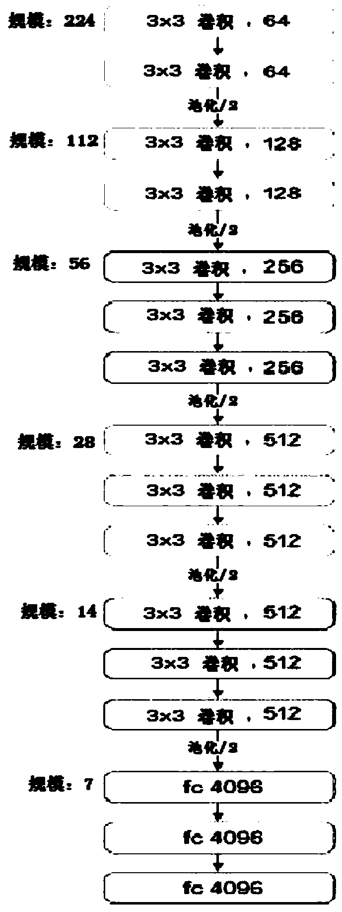
[0064] Use Caffe's open source deep learning computing framework to realize the progr...
PUM
 Abstract
Description
Claims
Application Information
 - R&D
- Intellectual Property
- Life Sciences
- Materials
- Tech Scout
- Unparalleled Data Quality
- Higher Quality Content
- 60% Fewer Hallucinations
Browse by: Latest US Patents, China's latest patents, Technical Efficacy Thesaurus, Application Domain, Technology Topic, Popular Technical Reports.
© 2025 PatSnap. All rights reserved.Legal|Privacy policy|Modern Slavery Act Transparency Statement|Sitemap|About US| Contact US: help@patsnap.com


