Needle tip enhanced Raman spectrum microscopic imaging device

A technique of tip-enhanced Raman and microscopic imaging, which is applied in measurement devices, Raman scattering, material analysis by optical means, etc., can solve problems such as the decrease of incident light field intensity and the need for further improvement of Raman spectral signal intensity, etc. To achieve the effect of local electric field enhancement, improve spatial resolution, and enhance strength

Pending Publication Date: 2020-04-07
SHENZHEN UNIV
View PDF0 Cites 6 Cited by
  • Summary
  • Abstract
  • Description
  • Claims
  • Application Information

AI Technical Summary

Problems solved by technology

However, the first method is only effective for transparent samples, and for non-transparent samples, the intensity of the incident light field will be reduced; although the second method can solve

Method used


Image

  • Needle tip enhanced Raman spectrum microscopic imaging device
  • Needle tip enhanced Raman spectrum microscopic imaging device
  • Needle tip enhanced Raman spectrum microscopic imaging device

Examples


Embodiment 1

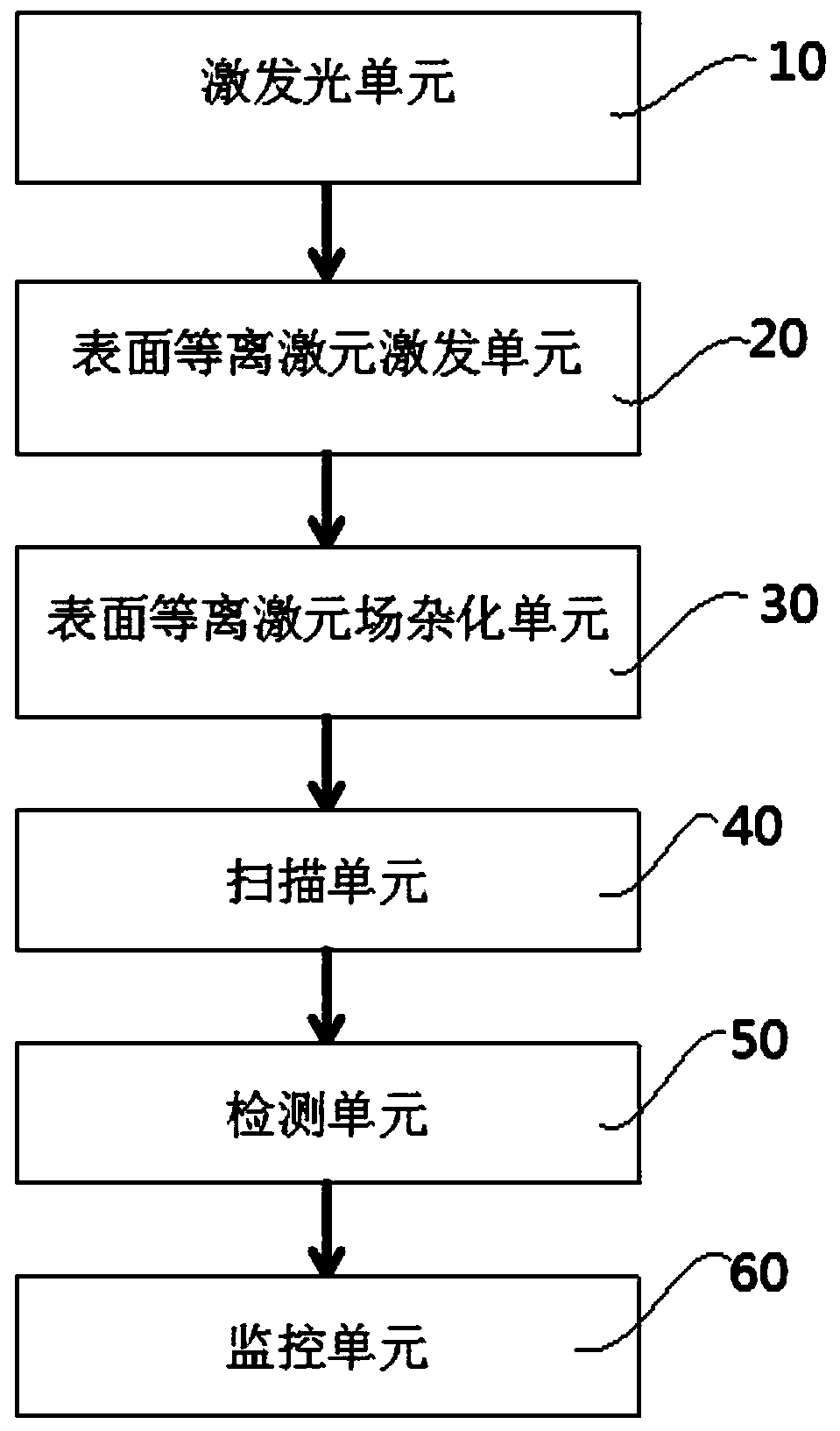
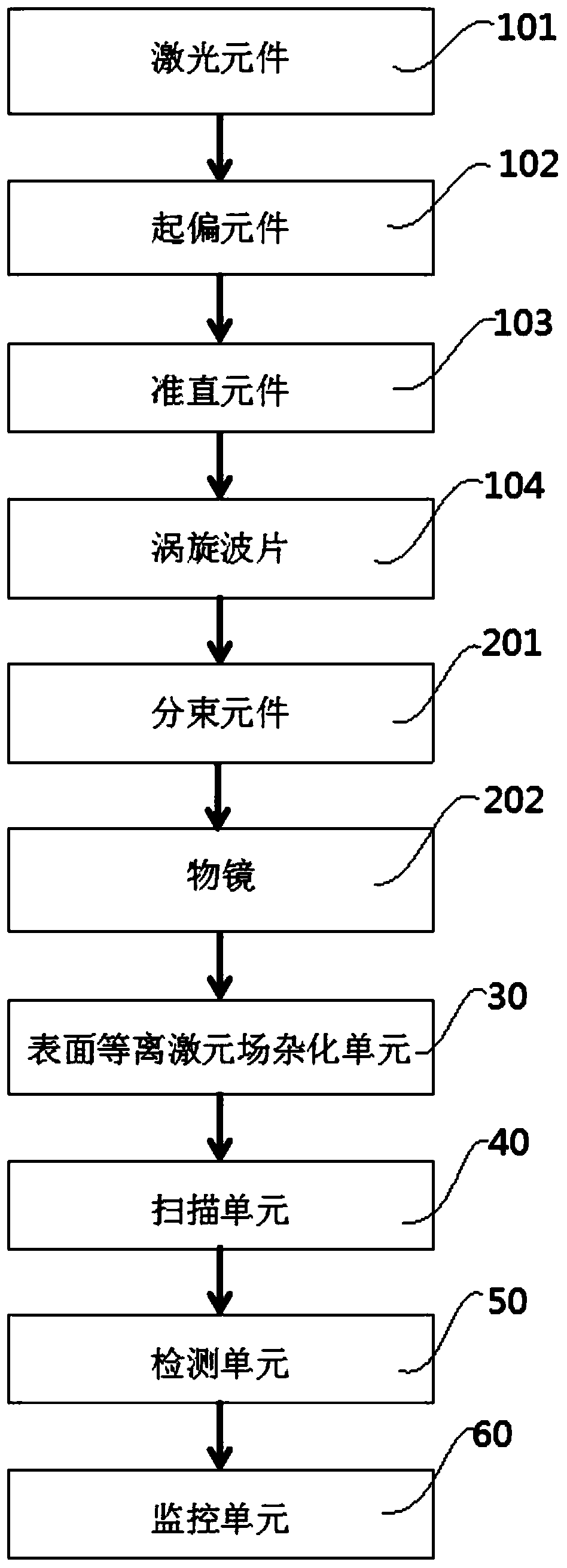
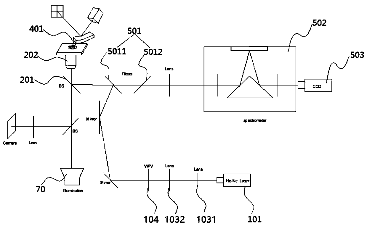
[0058] A needle-tip-enhanced Raman spectroscopy microscopic imaging device of this embodiment, such as Figure 1 to Figure 9 As shown, it includes an excitation light unit 10, a surface plasmon excitation unit 20, a surface plasmon field hybridization unit 30, an illumination unit 70, a scanning unit 40, a detection unit 50 and a monitoring unit 60; the excitation light unit 10 is used To generate radially polarized light beams, the generated radially polarized light beams are incident on the surface plasmon excitation unit 20; the surface plasmon excitation unit 20 is used to receive radially polarized light beams, irradiated by radially polarized light beams, and use The energy of the radially polarized light beam excites the surface plasmon light field; the scanning unit 40 includes a scanning probe 401; the scanning probe 401 is used to scan the sample to be tested, and the scanning probe is also used to interact with the surface plasmon The surface plasmon field hybridiza...


PUM

PropertyMeasurementUnit
Thicknessaaaaaaaaaa

Abstract

The invention provides a needle tip enhanced Raman spectrum microscopic imaging device. The device comprises: an exciting light unit for generating a radially polarized light beam; a surface plasmon excitation unit used for receiving the radially polarized light beam and exciting the radially polarized light beam to generate surface plasmon; and a scanning unit comprising a scanning probe, whereinthe scanning probe and the surface plasmon are hybridized to form a surface plasmon field hybridization unit, and the exciting light unit, the detection unit and the scanning unit are connected witha monitoring unit. Surface plasmon polaritons are generated through radial polarized light excitation and are focused to generate a surface plasmon polariton virtual probe, a gap structure formed between a scanning probe and a surface plasmon unit is hybridized with the surface plasmon to generate the surface plasmon field hybridization unit, so that a surface local electric field is enhanced, anenhanced Raman spectrum signal of a to-be-detected sample is obtained, and microscopic imaging of the sample is further realized by utilizing a Raman spectrum obtained by measurement.

Description

technical field [0001] The invention belongs to the technical field of microspectral imaging devices, in particular to a needle-tip enhanced Raman spectroscopic microimaging device. Background technique [0002] Raman spectroscopy is a common spectroscopic analysis method for detecting chemical bonds, symmetry or other chemical composition and structural information of sample molecules. Can provide fast, simple, repeatable and non-destructive qualitative and quantitative analysis. [0003] Tip-Enhanced Raman Spectroscopy (TERS) technology is a combination of Scanning Probe Microscopy (SPM) technology and Raman spectroscopy technology. TERS technology can meet the analysis requirements of chemical substances on the surface and interface in nanoscience and nanotechnology, with high spatial resolution and significant enhancement of molecular Raman signals. The principle is to control the Ag or Au tip with a radius of curvature of tens of nanometers at a very close distance (s...

Claims


Application Information

Patent Timeline
IPC IPC(8): G01N21/65
CPCG01N21/658
Inventor 张聿全黎佳星陈厚凯闵长俊袁小聪
Owner SHENZHEN UNIV
Why Eureka
Try Eureka