
Image processing method and system for assisting diagnosis of pneumonia

An image processing and pneumonia technology, applied in the field of medical imaging, can solve problems such as unfavorable conditions and limited clinical data, achieve stable diagnostic accuracy and improve clinical diagnostic efficiency

Active Publication Date: 2020-07-24
NANJING ANKE MEDICAL TECH CO LTD
View PDF7 Cites 0 Cited by
  • Summary
  • Abstract
  • Description
  • Claims
  • Application Information

AI Technical Summary

Problems solved by technology

Therefore, for emerging viral pneumonia, such as new coronavirus pneumonia, the available clinical data is very limited, which is not conducive to the application of traditional methods for treatment

Method used


Image

  • Image processing method and system for assisting diagnosis of pneumonia
  • Image processing method and system for assisting diagnosis of pneumonia
  • Image processing method and system for assisting diagnosis of pneumonia

Examples


Embodiment Construction

[0029] The present invention will be further described below in conjunction with the accompanying drawings.

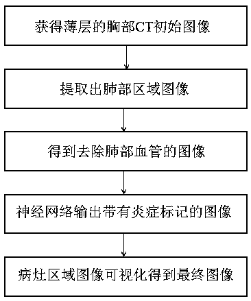
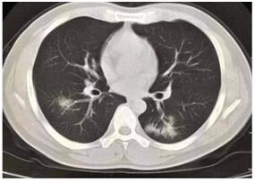
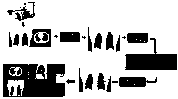
[0030] like Figure 1 to Figure 7 As shown, the present invention provides an image detection method for assisting the diagnosis of pneumonia, such as for diagnosing new coronary pneumonia, comprising steps:

[0031] A. Perform a low-dose CT scan on the patient to obtain thin-slice chest CT image data;

[0032] B. Segment the CT image and extract the image of the lung region;

[0033] C. Process the image of the lung area to remove the blood vessels in the lung;

[0034] D. Use the neural network to detect the image of the removed lung blood vessels, and distinguish the similarity between these areas and the new coronary pneumonia lesions;

[0035] E. Segment the lesion area in the image with inflammatory markers, and visualize the area for doctors to review.

[0036] In the present invention, step B can use the region growing + threshold segmentation method to com...


PUM


Abstract

The invention relates to the technical field of medical imaging, and discloses an image processing method and system for assisting pneumonia diagnosis. The method includes the steps of: obtaining a thin-layer chest CT initial image; performing segmentation processing on the initial image, and extracting an image of a lung region ; Process the image of the lung area to obtain an image without pulmonary blood vessels; use a neural network to detect the image without pulmonary blood vessels, and output an image with inflammatory markers. The training of the neural network includes using different training data sets and Pre-training and precise training of training objects; segment the lesion area in the image with inflammatory markers, and visualize the image of the lesion area to obtain the final image for doctors to review. Based on various image processing methods and neural network detection technology, the present invention can solve the neural network training problem of limited clinical pneumonia data, obtain stable diagnostic accuracy, and improve clinical diagnosis efficiency.

Description

technical field [0001] The invention relates to the technical field of medical imaging, in particular to an image processing method and system for assisting pneumonia diagnosis. Background technique [0002] Pneumonia is an acute respiratory infection that affects lung breathing. Pneumonia is easy to infect and has a relatively high mortality rate. Therefore, high requirements are placed on the rapid detection and diagnosis of pneumonia. At present, the diagnosis of pneumonia is generally based on the detection and diagnosis of physical signs and chest X-rays. However, it is not easy to determine the underlying cause of pneumonia in this way, because there is no standard that can clearly distinguish bacterial pneumonia from non-bacterial pneumonia. Pneumonia may not be visible on radiographs at an early stage because of the low contrast of radiographs and severe aliasing of tissue results, especially in the case of dehydration and other pulmonary diseases with a history of o...

Claims


Application Information

Patent Timeline
IPC IPC(8): G06T7/00G06T7/11G06T7/136G16H30/40G16H50/20A61B6/00
CPCA61B6/463A61B6/5217G06T7/0012G06T7/11G06T7/136G06T2207/10081G06T2207/20081G06T2207/20084G06T2207/30064G06T2207/30101G16H30/40G16H50/20
Inventor 曾凯何健郭桐冯亚崇杨奎吴小页
Owner NANJING ANKE MEDICAL TECH CO LTD
Learn More