Method for identifying parathyroid hyperplasia in ultrasonic image

A parathyroid and ultrasound image technology, applied in the field of image recognition, can solve the problems of cumbersome steps, large noise, small difference in image features, etc., and achieves the effect of no longer cumbersome intermediate process, reduced misjudgment rate, and easy identification.

Inactive Publication Date: 2020-04-24
BEIJING UNIV OF CHEM TECH
View PDF6 Cites 4 Cited by
  • Summary
  • Abstract
  • Description
  • Claims
  • Application Information

AI Technical Summary

Problems solved by technology

Therefore, SHPT seriously affects the quality of life of patients and shortens the survival period of patients.
[0003] Ultrasound examination is the most commonly used clinical diagnosis of SHPT at present, but there are many deficiencies in the ultrasonographic examination method: 1) Since ultrasonic imaging uses the acoustic signal reflected by human tissue for imaging, when the physical and chemical properties of different tissues are similar, compared with other

Method used


Image

  • Method for identifying parathyroid hyperplasia in ultrasonic image
  • Method for identifying parathyroid hyperplasia in ultrasonic image
  • Method for identifying parathyroid hyperplasia in ultrasonic image

Examples


Embodiment 1

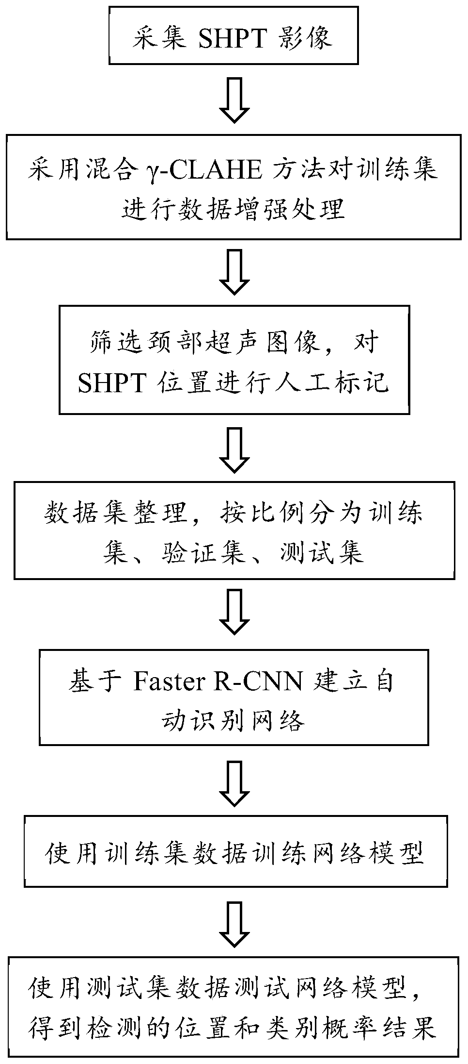
[0064] figure 1 As shown, the method includes:

[0065] S1. Acquire 1200 ultrasound images of parathyroid glands, and pay attention to collecting images of parathyroid glands in transverse section and longitudinal section during the acquisition process.

[0066] S2. Screening and dividing the obtained enhanced images, and labeling the data set;

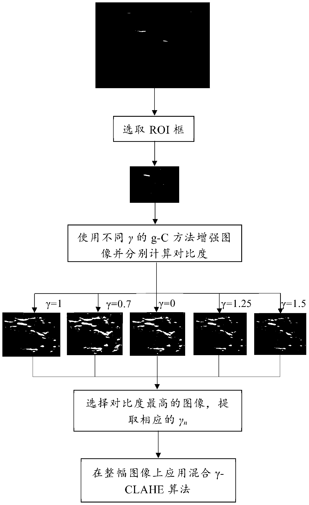
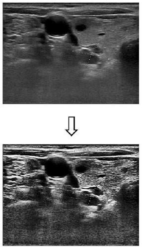
[0067] S3, using the method of combining CLAHE and gamma transformation to perform data enhancement processing on the training set;

[0068] S4. Train the Faster-RCNN network model that can identify parathyroid hyperplasia. Faster R-CNN is a combination of RPN (Region Proposal Network) and Fast R-CNN models, and Faster R-CNN has been improved in some networks. During training, alternately train RPN and Fast R-CNN networks;

[0069] S5. Use the trained Faster R-CNN model to test the new data set. For a test image, first run RPN to generate region proposals, and then project the region proposals to the conv feature map for subsequent...


PUM


Abstract

The invention discloses a method and a system for identifying parathyroid hyperplasia in an ultrasonic image. The method comprises the following steps: acquiring a training sample comprising the ultrasonic image and a corresponding diagnosis result, wherein the ultrasonic image is an image obtained after ultrasonic imaging is carried out on a hyperparathyroidism area of a patient; carrying out data enhancement processing on the training sample by adopting a mixed gamma-CLAHE method; inputting the enhanced training sample into a Faster R-CNN network to obtain a trained network model; and inputting the neck ultrasonic image to be recognized into the trained Faster R-CNN model to obtain a recognition result output by the trained model. The method is high in recognition accuracy and high in speed, can help doctors to quickly find out organ positions and judge organ possibility from the perspective of image recognition, and improves efficiency for the doctors.

Description

technical field [0001] The invention relates to the technical field of image recognition, in particular to an automatic recognition method in ultrasonic images of parathyroid hyperplasia. Background technique [0002] The prevalence of chronic kidney disease in Chinese adults is about 10-13%. There are 120 million kidney disease patients over the age of 18, and 20% of the patients will turn into uremia, that is, chronic renal failure (CRF). The total number of CRF patients in my country is about 1.38-2 million, and it is increasing at a rate of 100,000 per year. Secondary hyperparathyroidism (SHPT) is one of the most common adverse reactions in patients with chronic kidney disease (CKD). SHPT can also lead to serious conditions such as cardiomyopathy, hypertension, and hypercalcemia, which increases the risk of kidney stones. Therefore, SHPT seriously affects the quality of life of patients and shortens the survival period of patients. [0003] Ultrasound examination is t...

Claims


Application Information

Patent Timeline
IPC IPC(8): G06T7/11G06T7/136G06T5/40
CPCG06T5/40G06T2207/10132G06T2207/20081G06T2207/20084G06T2207/30096G06T7/11G06T7/136
Inventor 郭青郑睿之王华庆于明安高唱
Owner BEIJING UNIV OF CHEM TECH
Why Eureka
Try Eureka