Bone suppression image generation method and device, storage medium and electronic equipment

An image generation and bone suppression technology, applied in the field of image processing, can solve the problems of high cost and expensive equipment, and achieve the effect of reducing distortion and improving image clarity

Active Publication Date: 2020-04-28
INFERVISION MEDICAL TECH CO LTD
View PDF10 Cites 3 Cited by
  • Summary
  • Abstract
  • Description
  • Claims
  • Application Information

AI Technical Summary

Problems solved by technology

However, dual energy subtraction uses hardware equipment for bone suppression, but the equipment is expensive and the cost is high

Method used


Image

  • Bone suppression image generation method and device, storage medium and electronic equipment
  • Bone suppression image generation method and device, storage medium and electronic equipment
  • Bone suppression image generation method and device, storage medium and electronic equipment

Examples


Embodiment 1

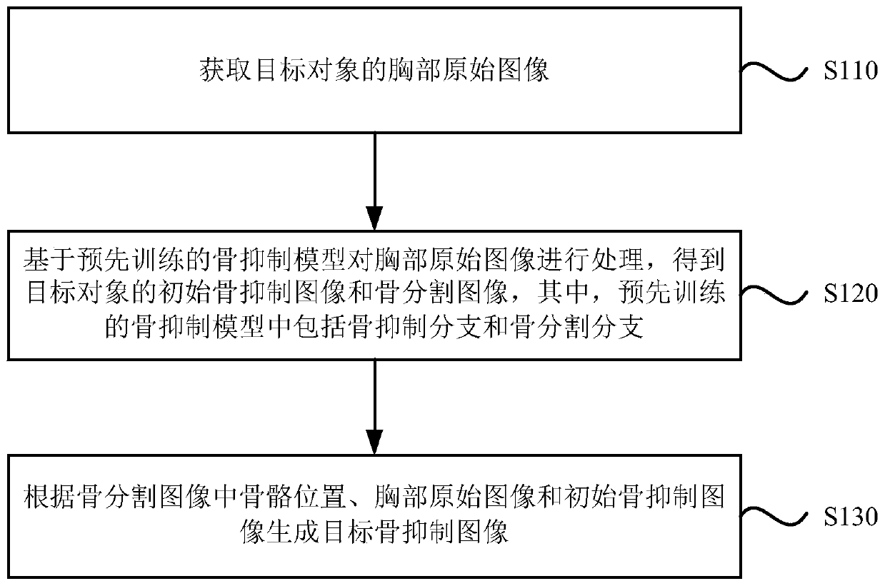
[0027] figure 1 It is a schematic flow chart of a method for generating a bone-suppressed image provided by Embodiment 1 of the present invention. This embodiment is applicable to the case of obtaining a high-precision bone-suppressed image. To execute, the apparatus may be realized by means of software and / or hardware, and the apparatus may be integrated into electronic equipment such as a server or a computer. The method specifically includes the following steps:

[0028] S110. Acquire an original chest image of the target object.

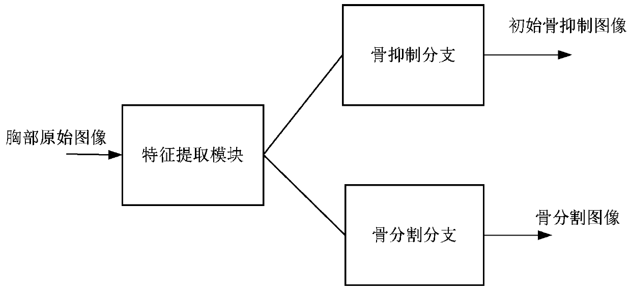
[0029] S120. Process the original breast image based on the pre-trained bone suppression model to obtain an initial bone suppression image and a bone segmentation image of the target object, wherein the pre-trained bone suppression model includes a bone suppression branch and a bone segmentation branch.

[0030] S130. Generate a target bone-suppressed image according to the bone position in the bone segmentation image, the original chest image,...

Embodiment 2

[0048] Figure 5 It is a schematic flowchart of a method for generating a bone-suppressed image provided by an embodiment of the present invention. On the basis of the above-mentioned embodiments, a training method for a bone-suppressed model is provided. The method specifically includes:

[0049] S210. Create an initial bone suppression model.

[0050] S220. Input the sample data into the initial bone suppression model to obtain a predicted bone suppression image and a predicted bone segmentation image.

[0051] S230. Train the initial bone suppression model based on the predicted bone suppression image and the standard bone suppression image, and train the initial bone suppression model based on the predicted bone segmentation image and the standard bone segmentation image.

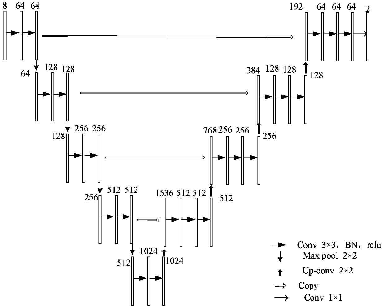
[0052] Among them, the structure of the initial bone suppression model can be as follows image 3 As shown, the initial bone suppression model is trained based on the pre-collected sample data and th...

Embodiment 3

[0068] Image 6 It is a schematic structural diagram of a device for generating a bone-suppressed image provided in Embodiment 3 of the present invention, and the device includes:

[0069] An original image acquisition module 310, configured to acquire an original image of the chest of the target object;

[0070]The image processing module 320 is configured to process the original chest image based on a pre-trained bone suppression model to obtain an initial bone suppression image and a bone segmentation image of the target object, wherein the pre-trained bone suppression model includes Osteoinhibition branch and bone segmentation branch;

[0071] A target bone-suppressed image determination module 330, configured to generate a target bone-suppressed image according to the bone position in the bone segmentation image, the original chest image and the initial bone-suppressed image.

[0072] Optionally, the bone segmentation image includes classification probabilities of each ...


PUM


Abstract

The invention discloses a bone suppression image generation method and a device, a storage medium and electronic equipment. The method comprises the steps of obtaining a chest original image of a target object; processing the chest original image based on a pre-trained bone suppression model to obtain an initial bone suppression image and a bone segmentation image of the target object, the pre-trained bone suppression model comprising a bone suppression branch and a bone segmentation branch; and generating a target bone suppression image according to the bone position in the bone segmentationimage, the chest original image and the initial bone suppression image. According to the method provided by the embodiment of the invention, the obtained target bone suppression image can eliminate the bony contour of the pixel points of the skeleton region, can reserve the original image information of the non-skeleton region, improves the image definition, and reduces the distortion of the bonesuppression model in the chest original image processing process.

Description

technical field [0001] Embodiments of the present invention relate to the technical field of image processing, and in particular to a method, device, storage medium, and electronic equipment for generating a bone-suppressed image. Background technique [0002] Digital radiography (digital radiography, DR) is a new digital imaging technology recently developed. It has similar uses to computer radiography (CR), but its basic principles and structures are different. DR is developed on the basis of digital fluorescence photography (digital fluorography, DF). It uses image intensifier tubes as information carriers to receive X-ray information that passes through the human body. After being collected by a video camera, it is converted into digital signals and then digitized. [0003] Chest X-rays are chest X-rays. Routine chest X-ray examinations include frontal, left anterior oblique (60°-65°), right anterior oblique (45°-55°) and left lateral views. Frontal chest radiography ca...

Claims


Application Information

Patent Timeline
Patent Type & Authority Applications(China)
IPC IPC(8): G06T5/50G06T7/10
CPCG06T5/50G06T2207/10124G06T2207/20081G06T2207/20084G06T2207/20192G06T2207/20221G06T2207/30008G06T7/10
Inventor 周越邹彤赵朝炜李新阳陈宽王少康
Owner INFERVISION MEDICAL TECH CO LTD
Why Eureka
Try Eureka