Image lesion area segmentation method and device and server

A lesion area and image technology, applied in the field of image recognition, can solve the problem of low accuracy of lesion area

Active Publication Date: 2020-04-28
SHENZHEN INST OF ADVANCED TECH CHINESE ACAD OF SCI
View PDF3 Cites 4 Cited by
  • Summary
  • Abstract
  • Description
  • Claims
  • Application Information

AI Technical Summary

Problems solved by technology

[0004] In view of this, an embodiment of the present invention provides a method, device and server for image lesion area segmentation to solve the problem of low accuracy in segmenting lesion areas in images

Method used


Image

  • Image lesion area segmentation method and device and server
  • Image lesion area segmentation method and device and server
  • Image lesion area segmentation method and device and server

Examples


Embodiment 1

[0042] Such as figure 1 As shown in FIG. 1 , it is a schematic flowchart of the image lesion region segmentation method provided by Embodiment 1 of the present invention. This embodiment is applicable to the application scenario of segmenting the lesion area in the image obtained by magnetic resonance scanning, and the method can be executed by an image lesion area segmentation device, which can be a server, an intelligent terminal, a tablet or a PC, etc.; in this application In the embodiment, the image lesion region segmentation device is used as the execution subject for illustration, and the method specifically includes the following steps:

[0043] S110, performing feature extraction on the image obtained by the magnetic resonance scan, to generate a feature image;

[0044] In medical pathological diagnosis, MRI is often used to scan the patient's body to obtain various parts of the body, so that doctors can determine the lesion in the patient's body by determining the l...

Embodiment 2

[0059] Such as figure 2 Shown is a schematic flow chart of the image lesion region segmentation method provided by Embodiment 2 of the present invention. On the basis of the first embodiment, this embodiment also provides a process of optimizing parameters in the image lesion region segmentation method, so as to further segment the accuracy of the lesion region in the image. The method specifically includes:

[0060] S210. Input preset image training data into the cascaded depth-separable convolutional layers in the encoder for feature extraction to generate first feature data;

[0061] In order to realize the determination of the lesion area in the image through the lesion area segmentation method of the deep learning image, it is necessary to input the preset image training data into the cascaded depth separable convolution layer in the encoder for feature extraction to generate the first feature data , to achieve the training of the encoder. Convolute each channel of th...

Embodiment 3

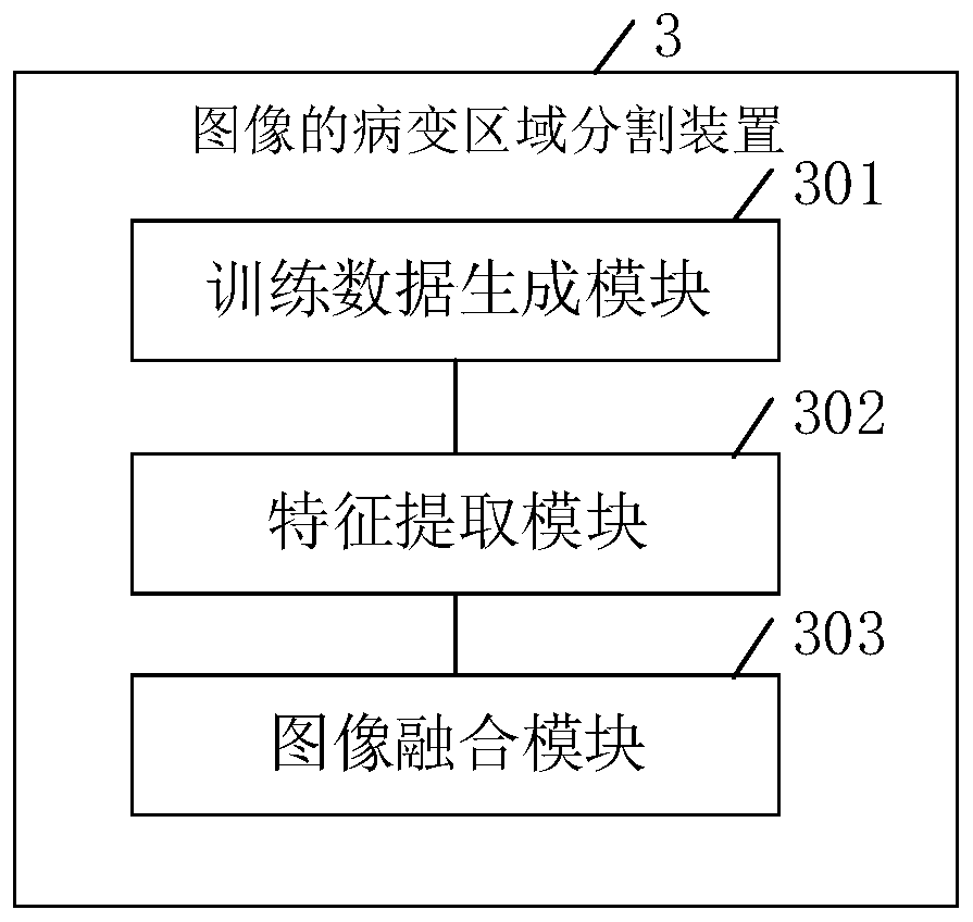
[0069] Such as image 3 Shown is the image lesion region segmentation device provided by Embodiment 3 of the present invention. On the basis of Embodiment 1 or 2, the embodiment of the present invention also provides an image lesion area segmentation device 3, which includes:

[0070] The feature extraction module 301 is used to perform feature extraction on the image obtained by the magnetic resonance scan to generate a feature image;

[0071] The similarity calculation module 302 is used to calculate the correlation between each pixel in the feature image through the feature similarity module to obtain a feature image containing similarity information;

[0072] The lesion area segmentation module 303 is configured to predict the lesion area in the feature image containing similarity information, and output an image of the lesion area.

[0073] In an implementation example, the feature similarity module includes:

[0074] An associated information calculation unit, configu...


PUM


Abstract

The invention belongs to the technical field of image recognition, and provides an image lesion area segmentation method and device, and a server, and the method comprises: carrying out feature extraction of an image obtained through magnetic resonance scanning, and generating a feature image; calculating the relevance between each pixel point in the feature image through a feature similarity module to obtain a feature image containing similarity information; and predicting a lesion area in the feature image containing the similarity information, and outputting a lesion area image. According to the embodiment of the invention, the problem of low accuracy of segmenting the lesion area in the image is solved.

Description

technical field [0001] The present invention relates to the technical field of image recognition, in particular to an image lesion area segmentation method, device and server. Background technique [0002] Magnetic resonance images of the brain can help specialists assess stroke lesions effectively and formulate effective treatment plans. In traditional methods, the segmentation of brain stroke lesion areas is usually done manually by professional radiologists slice by slice on magnetic resonance image slices, which is obviously time-consuming and highly subjective. At present, image segmentation methods mostly use methods based on deep learning such as convolutional neural networks to realize automatic segmentation of stroke lesion regions. In addition, dilated convolution operations and pyramid pooling structures are also introduced to obtain multi-scale feature maps for accurate prediction. In order to make full use of the correlation information between pixels, a netwo...

Claims


Application Information

Patent Timeline
Patent Type & Authority Applications(China)
IPC IPC(8): G06T7/11G06T7/00G06N3/04G06K9/62
CPCG06T7/0012G06T7/11G06T2207/10088G06T2207/30016G06N3/045G06F18/2135
Inventor 王珊珊郑海荣祁可翰刘新
Owner SHENZHEN INST OF ADVANCED TECH CHINESE ACAD OF SCI
Why Eureka
Try Eureka