Transmission electron microscope biological sample fixing tube for remote delivery

A technology of biological samples and transmission electron microscopy, applied in the preparation of test samples, discharge tubes, circuits, etc., can solve the problems of sample temperature reduction, influence on ultrastructure observation results, distortion, etc., and achieve the effect of real experimental results

Pending Publication Date: 2020-04-28
NANTONG UNIVERSITY
View PDF8 Cites 0 Cited by
  • Summary
  • Abstract
  • Description
  • Claims
  • Application Information

AI Technical Summary

Problems solved by technology

However, in the process of sending samples, there will be two unfavorable factors that will affect the follow-up observation results: First, the courier company will take care of each link in the transportation process, such as picking up items, scanning codes at outlets, loading, and checking at transfer stations. Transit, etc., will cause the centrifuge tube to be upside down and tilted due to the random placement of the package, so that the sample tissue block will adhere to the tube wall without fixative solution and leave the fixative solution environment for a long time, causing insufficient fixation of the sample. Observation results of ultrastructure; the second is that the samples are usually placed in 1.5ml or 2ml commercially available common centrifuge tubes during the mailing process. After the ice pack is in direct contact with the centrifuge tube, the temperature of the sample that is stuck to the wall and out of the fixative environment may be rapidly increased. Decrease and freeze, resulting in the formation of ice crystals inside the tissue cells, which will also seriously affect the maintenance of its ultrastructure, resulting in distortion of subsequent results

Method used


Image

  • Transmission electron microscope biological sample fixing tube for remote delivery

Examples


Embodiment Construction

[0023] The following will clearly and completely describe the technical solutions in the embodiments of the present invention with reference to the accompanying drawings in the embodiments of the present invention. Obviously, the described embodiments are only some of the embodiments of the present invention, not all of them. Based on the embodiments of the present invention, all other embodiments obtained by persons of ordinary skill in the art without making creative efforts belong to the protection scope of the present invention.

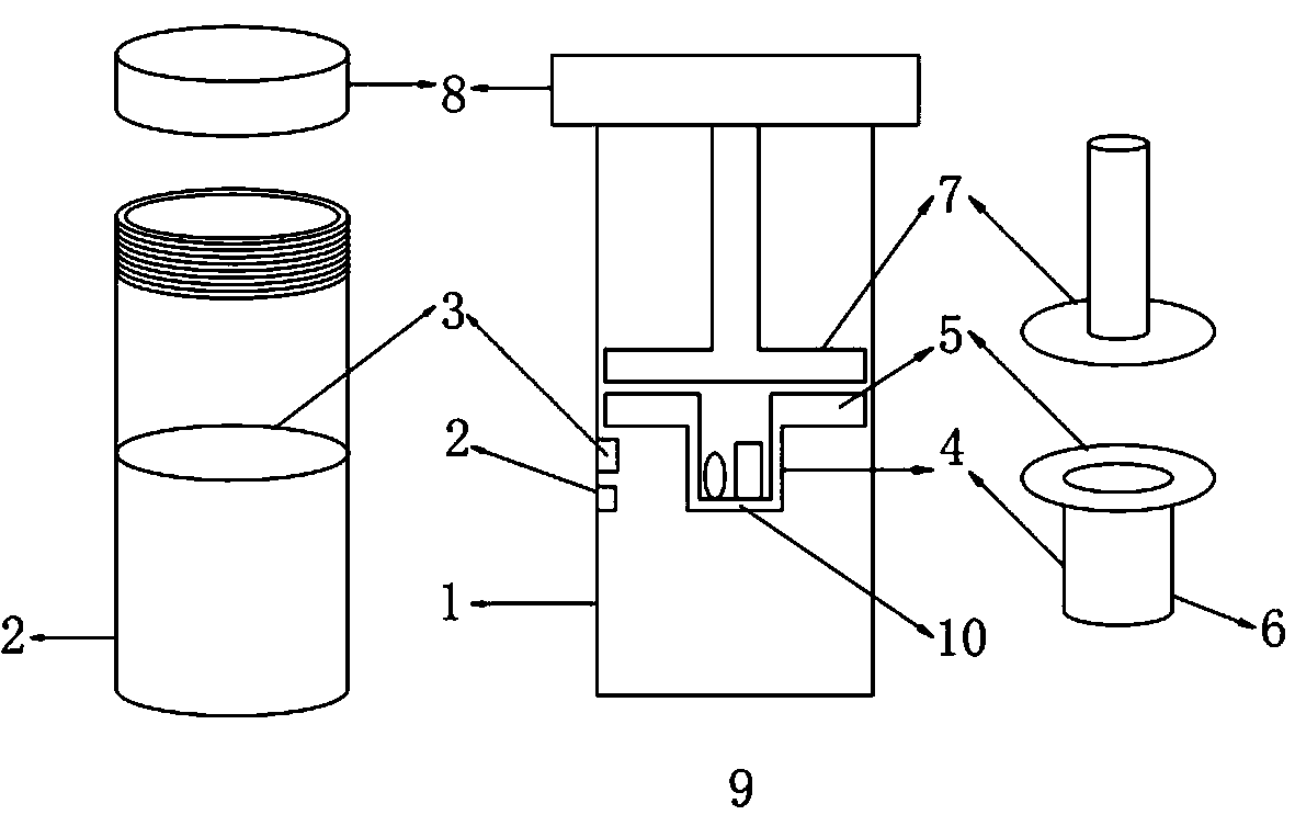
[0024] see figure 1 , the present invention provides a technical solution: a transmission electron microscope biological sample fixing tube for remote delivery, including a biological sample fixing tube body 9, a capacity tube A2 and a capacity tube B3 are arranged on the biological sample fixing tube body 9, and the capacity The inner center of the tube B3 is provided with a sample tank 4, and the top of the sample tank 4 is provided with a poro...


PUM

PropertyMeasurementUnit
thicknessaaaaaaaaaa
pore sizeaaaaaaaaaa
diameteraaaaaaaaaa

Abstract

The invention discloses a transmission electron microscope biological sample fixing tube for remote sending. The fixing tube comprises a biological sample fixing tube body, a sample tube blank tube isarranged on the biological sample fixing tube body; sample grooves, a porous plastic sheet with a connecting rod and a pipe cover are included, a transparent circular plastic tube serves as a hollowtube part; the tube orifice is of a screw structure; volume scale marks are marked on the outer surface of the tube wall; a circle of raised line is arranged near the middle lower position of the inner tube wall; the tube bottom is an arc-shaped round bottom or a sharp bottom; each sample groove is of a circular or square inverted cap-shaped structure; the depth of the sample groove is 3mm; the bottom of the sample groove is nest-shaped; and the sample groove is used for containing a sample. According to the transmission electron microscope biological sample fixing tube, the problem that the original ultrastructure of tissue cells cannot be fully maintained due to the fact that the sample is easily separated from the stationary liquid environment and adheres to the wall in the remote sending process of an existing tissue sample can be solved, the sample can be submerged in stationary liquid in the whole process, the ultrastructure of the sample cannot be affected, and the reality and reliability of subsequent experiment results are guaranteed.

Description

technical field [0001] The invention relates to a transmission biological sample fixing tube for remote delivery, in particular to a transmission electron microscope biological sample fixing tube for remote delivery. Background technique [0002] Electron microscopy technology, especially ultra-thin sectioning technology using transmission electron microscopy, is being more and more widely used in various fields of biomedicine, such as basic medicine, clinical pathological diagnosis, etc. In order to obtain objective, true and good follow-up electron microscope observation image results, TEM ultra-thin section technology involves many sample processing links (mainly including fixation, rinsing, dehydration, infiltration and embedding, trimming, semi-thin sectioning, tissue positioning, Ultra-thin sectioning, electronic staining, etc.), and has high requirements for sample sampling and subsequent processing steps. Among them, the pre-fixation after sample collection is an ex...

Claims


Application Information

Patent Timeline
Patent Type & Authority Applications(China)
IPC IPC(8): H01J37/20G01N1/30G01N1/42
CPCH01J37/20G01N1/30G01N1/42
Inventor 朱昌来贺小琴刘芳王莹洁张天一
Owner NANTONG UNIVERSITY
Why Eureka
Try Eureka