Artificial intelligence method and system for identifying retinal bleeding image

An artificial intelligence and retinal technology, applied in the field of medical image processing, can solve the problems of visual function damage, low possibility, lack of fundus doctors, etc., and achieve the goal of reducing irreversible damage, accurate screening, and improving screening efficiency. Effect

Pending Publication Date: 2020-05-19
ZHONGSHAN OPHTHALMIC CENT SUN YAT SEN UNIV
View PDF5 Cites 2 Cited by
  • Summary
  • Abstract
  • Description
  • Claims
  • Application Information

AI Technical Summary

Problems solved by technology

The inspection process is time-consuming and laborious. For the current inspection of retinal hemorrhage, there are mainly the following problems:
[0003] 1. The examination of retinal hemorrhage can only be carried out in ophthalmology specialists, especially in the initial stage of the disease, the symptoms are often not obvious, and patients often miss the best treatment time because of this. For peripheral hemorrhage, patients and professionals are often required. Experienced ophthalmologists can only find out when the two cooperate
[0004] 2. After the retinal hemorrhage is detected, it is necessary to determine the urgency of the patient's need for treatment. Often some retinal hemorrhage, such as macular hemorrhage, will cause a sharp decrease in vision. This often requires professional ophthalmologists to guide, but community hospitals or level Low-level general hospitals and physical examination centers do not have professional ophthalmologists, and cannot make timely judgments on the degree of urgency
[0005] 3. Although wide-area fundus images can cover almost the entire circumference of the retina, accurate interpretation of this fundus image requires professionally trained ophthalmologists and a long period of experience accumulation
This often leads to delays in diagnosis and treatment and irreversible damage to vision, and severe cases lead to irreversible permanent blindness, causing serious burdens to individuals, families and society
However, detailed inspection of retinal hemorrhage is time-consuming and requires professional and experienced ophthalmologists, and the possibility of large-scale population screening is low. Therefore, rapid and effective automatic screening of retinal hemorrhage can be effectively used here Diagnose it in the early stage of the disease, and automatically judge the urgency of the bleeding, so that the patient can correctly understand his condition. If the situation is urgent, he should immediately go to the ophthalmology department of the hospital for treatment to avoid irreversible damage to the visual function.

Method used


Image

  • Artificial intelligence method and system for identifying retinal bleeding image
  • Artificial intelligence method and system for identifying retinal bleeding image
  • Artificial intelligence method and system for identifying retinal bleeding image

Examples


Embodiment 1

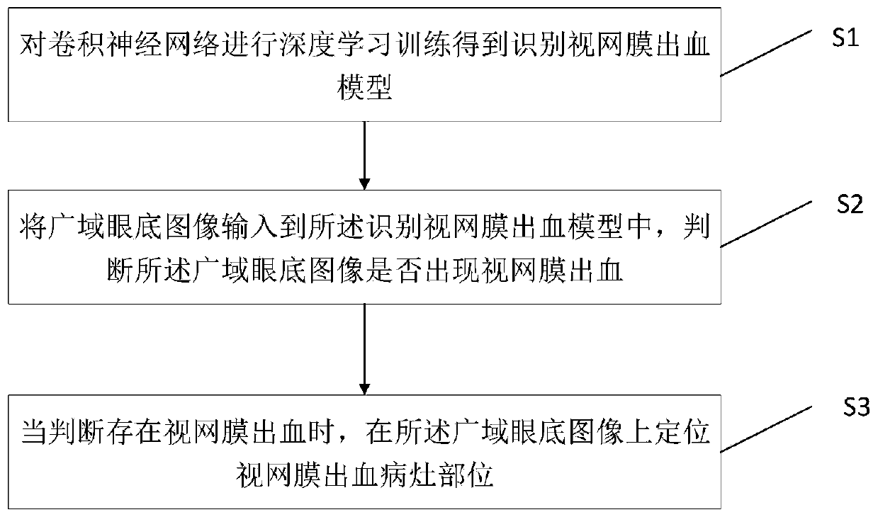
[0061] Such as figure 1 as shown, figure 1 It is a step diagram of an artificial intelligence method for recognizing retinal hemorrhage images according to the present invention, including the following steps:

[0062] S1. Perform deep learning training on the convolutional neural network to obtain a retinal hemorrhage recognition model;

[0063] S2. Input the wide-area fundus image into the model for identifying retinal hemorrhage, and judge whether there is retinal hemorrhage in the wide-area fundus image;

[0064] S3. When it is judged that there is retinal hemorrhage, locate the lesion site of retinal hemorrhage on the wide-area fundus image.

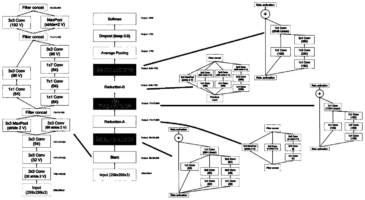
[0065] In the embodiment of the present invention, the accurate and efficient screening of retinal hemorrhage is realized. The specific implementation process of the artificial intelligence method for identifying retinal hemorrhage images is as follows: on the premise of having a large number of clearly classified wide-area fundus...

Embodiment 2

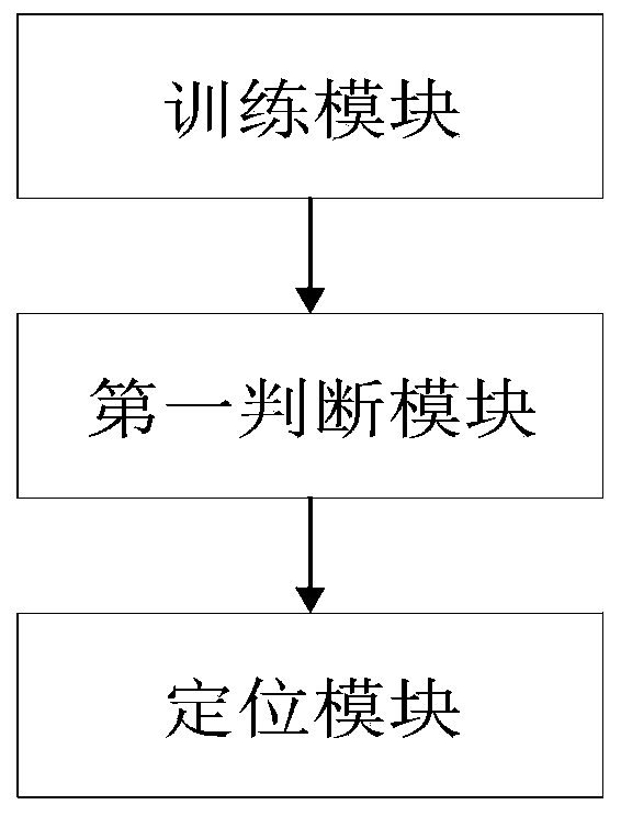
[0096] Such as image 3 as shown, image 3 It is a structural diagram of an artificial intelligence system for recognizing retinal hemorrhage images according to the present invention, and its system includes:

[0097] The training module is used to perform deep learning training on the convolutional neural network to obtain a retinal hemorrhage recognition model;

[0098] The first judging module is used to input the wide-area fundus image into the model for identifying retinal hemorrhage, and judge whether there is retinal hemorrhage in the wide-area fundus image;

[0099] The positioning module is configured to locate retinal hemorrhage lesions on the wide-area fundus image when it is judged that there is retinal hemorrhage.

[0100] In the embodiment of the present invention, accurate and efficient screening of retinal hemorrhage is realized. The implementation of the artificial intelligence system for recognizing retinal hemorrhage images is as follows: on the premise o...


PUM


Abstract

The invention relates to the field of medical image processing, in particular to an artificial intelligence method and a system for identifying a retinal hemorrhage image, and the method comprises thefollowing steps: carrying out the deep learning training of a convolutional neural network, and obtaining a retinal hemorrhage recognition model; inputting the wide-area fundus image into the retinalhemorrhage recognition model, and judging whether retinal hemorrhage occurs in the wide-area fundus image or not; and when it is judged that retinal bleeding exists, positioning a retinal bleeding focus part on the wide-area fundus image. According to the method, the retina of the wide-area fundus image is analyzed by means of the sensitivity and accuracy of artificial intelligence deep learning,so that early screening of retinal bleeding is more accurate, more intelligent and more portable, the screening efficiency is improved, and irreversible damage to people caused by retinal bleeding isreduced.

Description

technical field [0001] The present invention relates to the field of medical image processing, in particular to an artificial intelligence method and system for recognizing retinal hemorrhage images. Background technique [0002] The inspection process of retinal hemorrhage often requires the patient to dilate the pupil, which takes about 10 minutes, and then requires a professional ophthalmologist to conduct a full retinal exploration through an ophthalmoscope and determine the urgency of the hemorrhage requiring treatment. The inspection process is time-consuming and laborious. For the current inspection of retinal hemorrhage, there are mainly the following problems: [0003] 1. The examination of retinal hemorrhage can only be carried out in ophthalmology specialists, especially in the initial stage of the disease, the symptoms are often not obvious, and patients often miss the best treatment time because of this. For peripheral hemorrhage, patients and professionals are ...

Claims


Application Information

Patent Timeline
Patent Type & Authority Applications(China)
IPC IPC(8): G06T7/00G06K9/62G06N3/04
CPCG06T7/0012G06T2207/30041G06T2207/20081G06T2207/20084G06T2207/30096G06V2201/03G06N3/045G06F18/241
Inventor 林浩添李中文郭翀林铎儒
Owner ZHONGSHAN OPHTHALMIC CENT SUN YAT SEN UNIV
Why Eureka
Try Eureka