
Method for automatically estimating choroid thickness

A choroidal, automatic technology, applied in the field of medical image processing, can solve problems such as poor flexibility, achieve the effects of improving accuracy, resisting noise interference, and simple calculation

Active Publication Date: 2020-07-03
WEST CHINA HOSPITAL SICHUAN UNIV
View PDF9 Cites 0 Cited by
  • Summary
  • Abstract
  • Description
  • Claims
  • Application Information

AI Technical Summary

Problems solved by technology

However, this method needs to collect a large number of accurately labeled samples for training, and has poor flexibility in actual use.

Method used


Image

  • Method for automatically estimating choroid thickness
  • Method for automatically estimating choroid thickness
  • Method for automatically estimating choroid thickness

Examples


Embodiment Construction

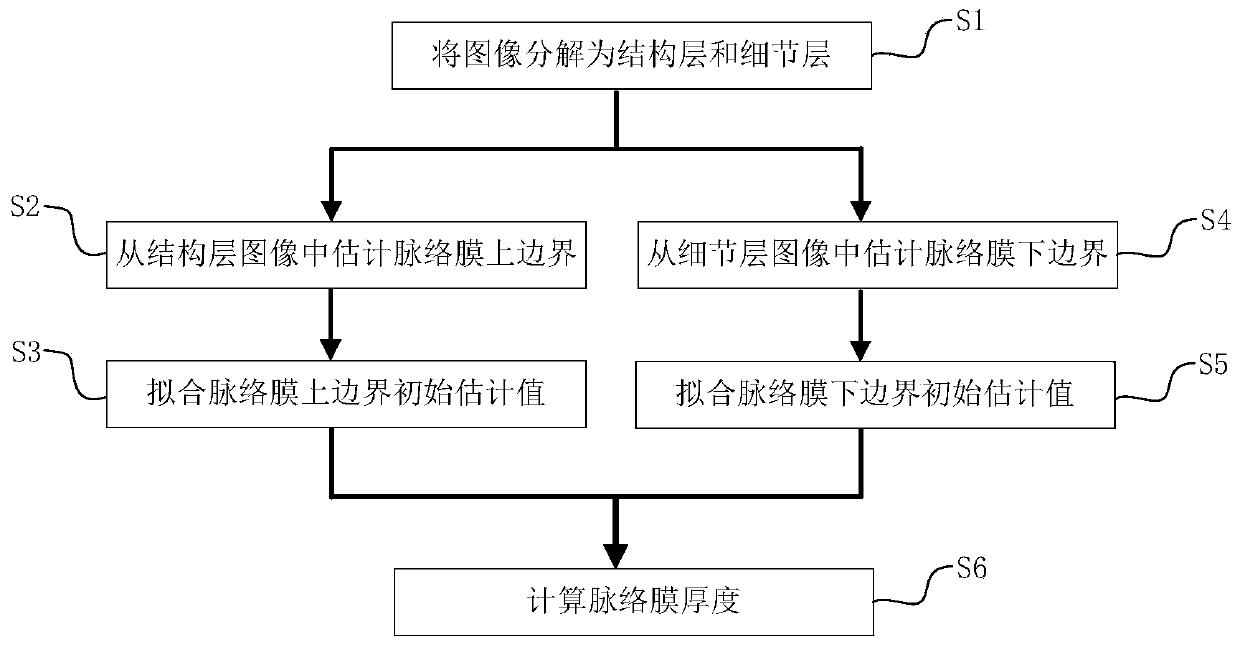
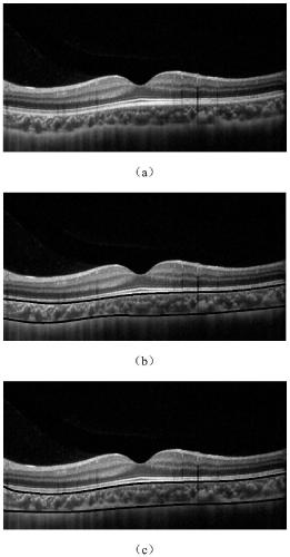
[0028] A real OCT image from West China Hospital of Sichuan University was used as the implementation object. The image size was 400×765, the format was 8-bit jpg format grayscale image, and the scale relationship was 4 μm / pixel. The specific calculation process is as follows figure 1 As shown, the specific process is as follows:

[0029] S1. Decompose the input image into a structure layer image and a detail layer image: set the regularization parameter to 0.2, decompose the input grayscale image based on the full variation model, and obtain the decomposed structure layer image and detail layer image;

[0030] S2. Estimate the initial position of the upper border of the choroid: for the structural layer image obtained by image decomposition in step S1, take the 100th column of the image as an example, the maximum value of the pixel in the 100th column of the image is 0.7031, so the threshold value is set to 0.6328 (that is, 0.7031 multiplied by 0.9); then find the pixels who...


PUM


Abstract

The invention discloses a method for automatically estimating choroidal thickness, which is applied to a medical image processing technology and aims at solving the problems of high complexity, largemanual estimation error and low estimation efficiency of the existing automatic choroidal thickness estimation algorithm. The method comprises the following steps: firstly, carrying out image decomposition on a fundus OCT image to obtain a structural layer image and a detail layer image; estimating the suprachoroidal boundary from the structural layer image; secondly, estimating a choroidal lowerboundary from the detail layer image; and finally, calculating the choroid thickness according to the estimated upper choroid boundary and the estimated lower choroid boundary. The method provided bythe invention has the characteristics of simple calculation, good effect and the like, and is very suitable for automatically estimating the thickness of the choroid of the fundus OCT image clinically.

Description

technical field [0001] The invention belongs to medical image processing technology, in particular to a technology for segmenting choroid from fundus optical coherence tomography (OCT) images and estimating choroid thickness. Background technique [0002] In fundus OCT images, changes in choroidal thickness can provide very important reference information for fundus lesions (such as choroidal diseases), as well as the occurrence, development, prevention and control of myopia. However, due to the complex structure of the fundus and the limited imaging quality of OCT technology, OCT images obtained from machine equipment often have low contrast and high noise. Therefore, in actual clinical applications, doctors often need to manually mark and measure the choroidal thickness. Manual measurement of choroidal thickness is not only time-consuming and laborious, but also there are errors among different doctors, which brings a lot of inconvenience to the follow-up diagnosis and tr...

Claims


Application Information

Patent Timeline
Patent Type & Authority Applications(China)
IPC IPC(8): G06T7/00G06T7/13G06T7/60
CPCG06T7/0012G06T7/13G06T7/60G06T2207/10101G06T2207/30041
Inventor 王将栏刘陇黔边思予何霄
Owner WEST CHINA HOSPITAL SICHUAN UNIV
Learn More