Method and device for predicting dose volume histogram of organ-at-risk of radiotherapy plan

A prediction method and histogram technology, applied in the field of medical radiation therapy, can solve the problems of not covering the characteristics of the patient's anatomical structure, reducing the accuracy and scope of clinical application, etc.

Pending Publication Date: 2020-07-28
戴建荣 +1
View PDF5 Cites 2 Cited by
  • Summary
  • Abstract
  • Description
  • Claims
  • Application Information

AI Technical Summary

Problems solved by technology

However, in clinical practice, the above methods require manual feature extraction, and these features cannot cover all the anatomical characteristics of patients, especially for complex tumor types (head and neck tumors), which reduces the accuracy and scope of clinical application

Method used


Image

  • Method and device for predicting dose volume histogram of organ-at-risk of radiotherapy plan
  • Method and device for predicting dose volume histogram of organ-at-risk of radiotherapy plan
  • Method and device for predicting dose volume histogram of organ-at-risk of radiotherapy plan

Examples


Embodiment 1

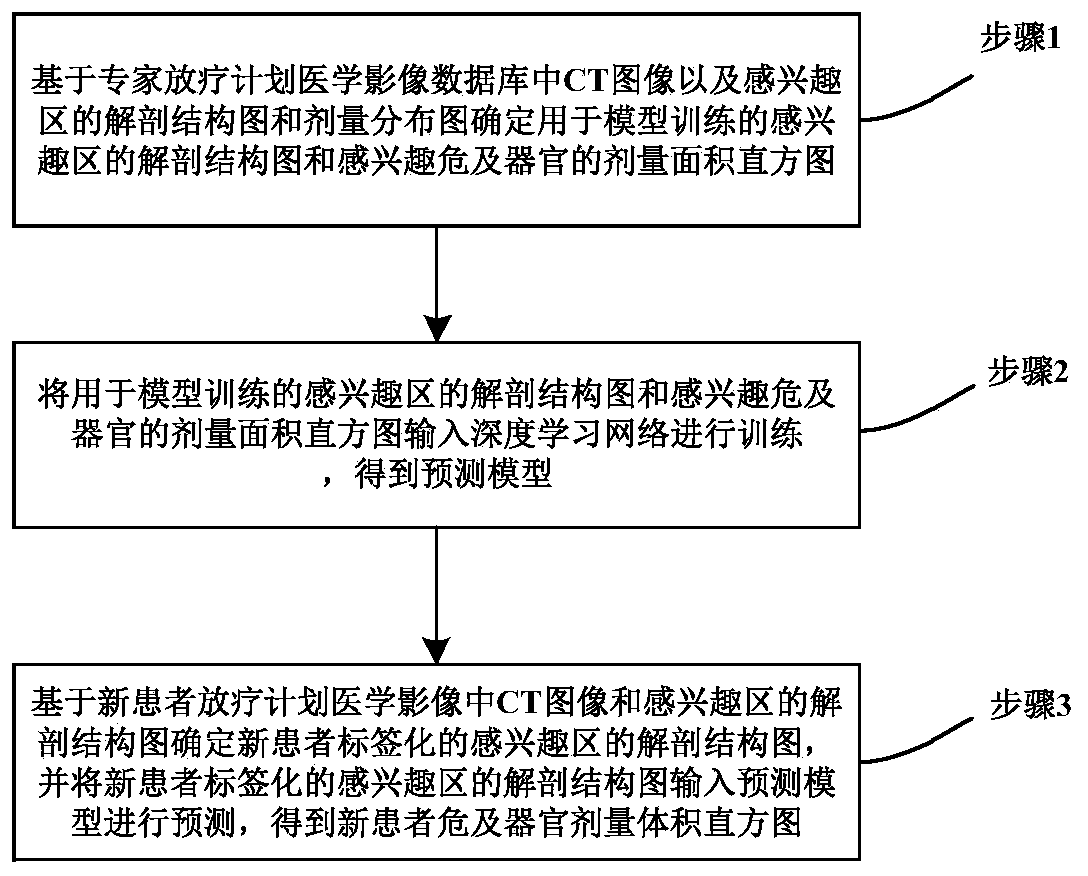
[0076] Embodiment 1 of the present invention provides a method for predicting the dose volume histogram (Dose Volume Histogram, DVH) of organs at risk in radiotherapy planning, and the specific flow chart is as follows figure 1 As shown, the specific process is as follows:

[0077] Step 1: Based on the CT images in the expert radiotherapy planning medical image database and the anatomical structure map and dose distribution map of the region of interest (body contour-target region-organ-at-risk), determine the anatomical structure map and sensory diagram of the region of interest used for model training. Dose Area Histogram (DAH) for organs at risk of interest;

[0078] In step 1, a medical image database of expert radiotherapy plans is established. The basis for selection is that the radiotherapy plan data is complete and the anatomical structure of the region of interest is clearly outlined. Senior physicists and radiotherapy doctors will confirm together to ensure the sel...

Embodiment 2

[0132] Based on the same inventive concept, Embodiment 2 of the present invention also provides a device for predicting the dose volume histogram of organs at risk in radiotherapy planning, such as figure 2 As shown, the functions of each component are described in detail below:

[0133] The determination module is used to determine the anatomical structure diagram of the region of interest and the dose area histogram of the organ at risk of interest for model training based on the CT image in the expert radiotherapy planning medical image database and the anatomical structure diagram and dose distribution diagram of the region of interest ;

[0134] The modeling module is used to input the anatomical structure map of the region of interest used for model training and the dose area histogram of the organ at risk of interest into the deep learning network for training to obtain a prediction model;

[0135] The prediction module is used to determine the anatomical structure ma...

Embodiment 3

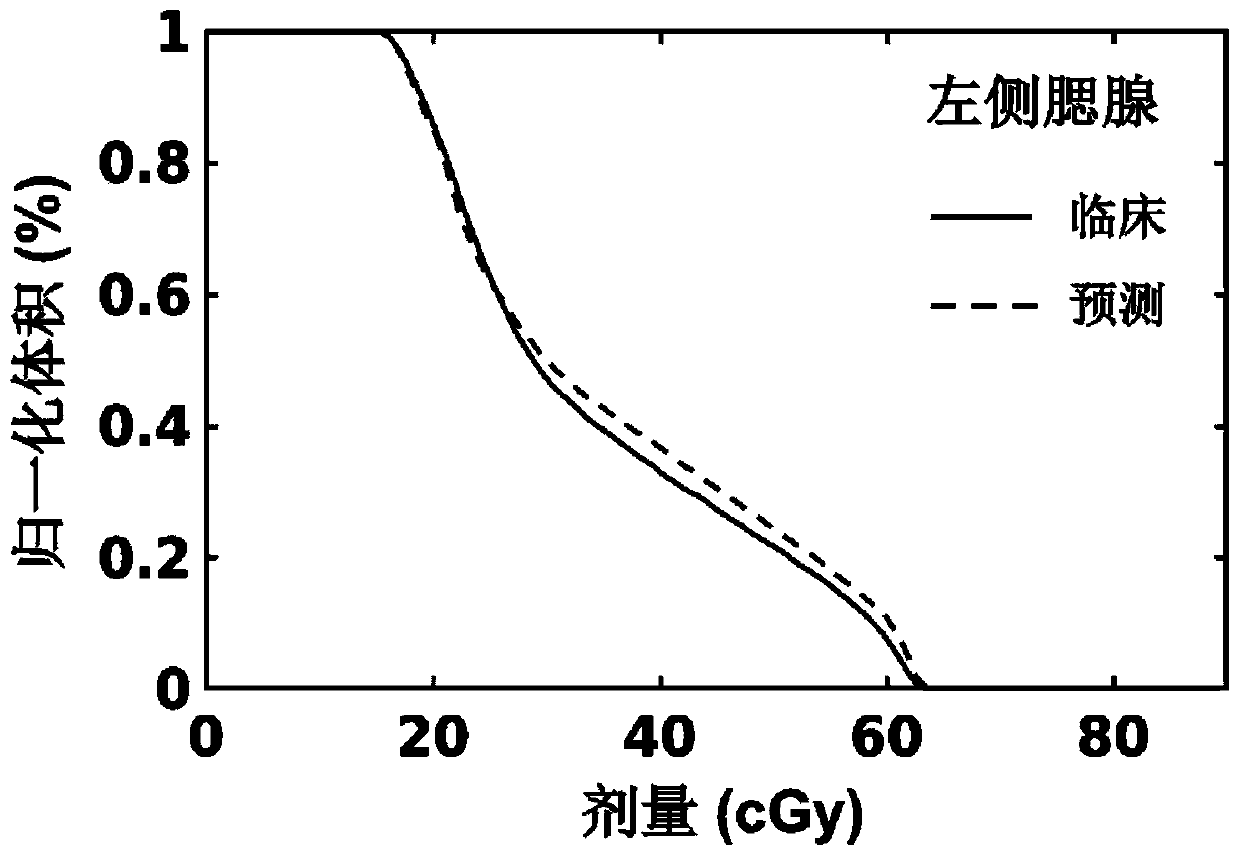
[0183] Embodiment 3 of the present invention takes nasopharyngeal carcinoma as an example to introduce the method of predicting the dose-volume histogram of organs at risk in the radiotherapy plan, and completes the DVH prediction of new patients to realize the process of automatically setting the initial objective function of the reverse plan and the plan quality inspection, specifically including the following steps :

[0184] (T1) Establish an expert database for radiotherapy planning for nasopharyngeal carcinoma, including the following:

[0185] (T1.1) Collect the radiotherapy planning data of 190 TOMO nasopharyngeal carcinoma patients admitted to the Cancer Hospital of the Chinese Academy of Medical Sciences from 2014 to 2017, among which 136 cases were randomly selected as the training data set, 34 cases were used as the verification data set, and 20 cases were used as the test data set;

[0186] (T1.2) For each radiotherapy plan, it includes CT images, anatomical stru...


PUM


Abstract

The invention provides a method and a device for predicting a dose volume histogram of an organ-at-risk of a radiotherapy plan. The method comprises the following steps: determining an anatomical structure diagram of a region-of-interest and a dose area histogram of an organ-at-risk for model training; determining a prediction model; and inputting the labeled anatomical structure diagram of the region-of-interest of a new patient into the prediction model, and predicting to obtain the organ-at-risk dose volume histogram of the new patient. According to the invention, the method and the deviceare short in prediction time consumption, high in accuracy and suitable for target regions with various prescription doses and patients with various different organ-at-risk anatomical structures, arenot limited to tumor types, can predict different tumor types, are wide in clinical application range, and can achieve automatic setting and plan quality inspection of the individualized reverse planinitial objective function, improve the radiotherapy plan design quality, shorten the design time and solve the problems of time consumption and large plan quality difference of accurate radiotherapyplan design.

Description

technical field [0001] The invention relates to the technical field of medical radiation therapy, in particular to a method and device for predicting a dose volume histogram of an organ at risk in a radiotherapy plan. Background technique [0002] Radiation therapy (radiotherapy, RT) is an important means of tumor treatment, more than 50% of tumor patients need to receive palliative or radical radiotherapy, including intensity-modulated radiation therapy (IMRT), fixed-field intensity-modulated radiation therapy (FB-IMRT), Volume modulated radiotherapy (VMAT) and tomospheric tomotherapy (TOMO) are currently the mainstream techniques for tumor radiotherapy. The purpose of the IMRT plan is to ensure that the tumor target area receives sufficient irradiation dose, and at the same time reduce the exposure dose to normal tissues and organs at risk around the target area as much as possible. However, the design of IMRT plans is complex and time-consuming, relying on the designer’s...

Claims


Application Information

Patent Timeline
Patent Type & Authority Applications(China)
IPC IPC(8): G16H50/70G16H50/20G16H30/20G06T7/00G06F16/583
CPCG16H50/70G16H50/20G16H30/20G06T7/0012G06F16/583G06T2207/10081G06T2207/20081G06T2207/20084
Inventor 戴建荣刘志强
Owner 戴建荣
Why Eureka
Try Eureka