Mammary gland image recognition method and device

An image recognition and mammary gland technology, applied in the medical field, can solve the problems of low diagnostic efficiency and easy misdiagnosis of mammary gland image recognition, and achieve the effect of improving efficiency and accuracy

Pending Publication Date: 2020-08-18
THE AFFILIATED HOSPITAL OF GUIZHOU MEDICAL UNIV
View PDF1 Cites 1 Cited by
  • Summary
  • Abstract
  • Description
  • Claims
  • Application Information

AI Technical Summary

Problems solved by technology

[0004] In the existing technology, the efficiency of breast image recognition and diagnosis is low, and the diagnosis results are too subjective, which is prone to misdiagnosis, and secondly, it is convenient to realize remote data transmission. Therefore, it is urgent to develop a method and device for breast image recognition

Method used


Image

  • Mammary gland image recognition method and device

Examples


Embodiment Construction

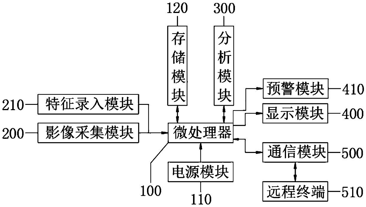
[0023] In order to make the above objects, features and advantages of the present invention more comprehensible, specific implementations of the present invention will be described in detail below in conjunction with the accompanying drawings.

[0024] In the following description, a lot of specific details are set forth in order to fully understand the present invention, but the present invention can also be implemented in other ways different from those described here, and those skilled in the art can do it without departing from the meaning of the present invention. Similarly generalized, the present invention is therefore not limited by the specific embodiments disclosed below.

[0025] Secondly, the present invention is described in detail in conjunction with schematic diagrams. When describing the implementation of the present invention in detail, for the convenience of explanation, the cross-sectional view showing the device structure will not be partially enlarged accor...


PUM


Abstract

The invention belongs to the technical field of medical treatment, and particularly relates to a mammary gland image recognition method which comprises the following steps: acquiring a to-be-detectedmammary gland image, and detecting mammary glands by using a detection device, the detection method comprising mammary gland molybdenum target X-ray photography, B-ultrasonic, CT, MRI and nuclide imaging; and acquiring a lump image according to mammary gland development, inputting the lump image into a processing center, carrying out data analysis on a mammary gland lump, obtaining a typing resultto which the mammary gland lump belongs according to the lump in the image, and meanwhile, judging the mammary gland position corresponding to the mammary gland lump according to the mammary gland lump. A mammary gland image recognition device comprises a microprocessor, an image acquisition module, an analysis module, a display module and a communication module, and the microprocessor is in electrical input connection with a power module and the image acquisition module. The accuracy of mammary gland diagnosis is improved, the diagnosis efficiency is improved, and meanwhile the comprehensiveeffect of remote transmission of data is achieved.

Description

technical field [0001] The invention relates to the field of medical technology, in particular to a method and device for breast image recognition. Background technique [0002] Breast cancer is a malignant tumor with a high incidence in women. Public data show that breast cancer accounts for 23% of all cancer cases and 14% of cancer-related deaths in women worldwide. Early screening can effectively reduce the incidence and mortality of breast cancer. Mammography is a relatively advanced examination method with good definition and high contrast, and can reliably identify benign lesions and malignant tumors of the breast. Lumps are a major manifestation of breast disease on mammograms. Due to the non-rigidity of breast tissue and the transformation of different viewing angles, the variability of shape, size, edge, and texture of lesions, the visual interference of normal tissue, and the large amount of film reading by doctors, it is easy to cause misdiagnosis and missed dia...

Claims


Application Information

Patent Timeline
Patent Type & Authority Applications(China)
IPC IPC(8): A61B8/08A61B6/00A61B5/00
CPCA61B8/5261A61B6/5247A61B5/0035A61B6/502A61B8/0825A61B5/004A61B5/4312
Inventor 柴鑫任宇
Owner THE AFFILIATED HOSPITAL OF GUIZHOU MEDICAL UNIV
Why Eureka
Try Eureka