Novel multi-modal fusion auxiliary diagnosis method based on rectal cancer imaging omics research

A radiomics, auxiliary diagnosis technology, applied in the field of medical image recognition and processing, can solve the problem of less application

Pending Publication Date: 2020-08-28
JILIN UNIV FIRST HOSPITAL
View PDF9 Cites 5 Cited by
  • Summary
  • Abstract
  • Description
  • Claims
  • Application Information

AI Technical Summary

Problems solved by technology

However, its application in

Method used


Image

  • Novel multi-modal fusion auxiliary diagnosis method based on rectal cancer imaging omics research
  • Novel multi-modal fusion auxiliary diagnosis method based on rectal cancer imaging omics research
  • Novel multi-modal fusion auxiliary diagnosis method based on rectal cancer imaging omics research

Examples


Embodiment 1

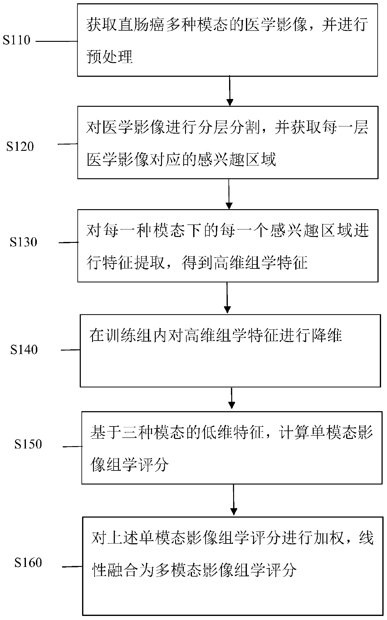
[0081] Step 1. Collect rectal cancer data. A patient has MRI T2WI, DWI (diffusion weighted imaging, a new MR imaging technology) sequence, and CT venous phase thick-layer image. There are three modal data in total. The collected data is divided into a training set and a verification set in a ratio of 7:3;

[0082] Step 2. Firstly, a radiologist segmented the layers of interest (VOIs) layer by layer on the T2WI, DWI and (enhanced CT) CE CT images respectively, and then the second radiologist independently randomized In each modality, 30 patient images were selected for layer-by-layer segmentation, and the VOI was drawn twice according to the same steps after a one-week interval. Both radiologists were blinded to the clinicopathological and other imaging findings.

[0083] Step 3. Extract radiomics features from the three modalities VOIs of T2WI, DWI and CE-CT respectively. Each sequence has 396 features, for a total of 1188 features.

[0084] Step 4, omics feature types incl...


PUM


Abstract

The invention provides a novel multi-modal fusion auxiliary diagnosis method based on rectal cancer imaging omics research. The method comprises the following steps of: 1, acquiring medical images ofmultiple modes of rectal cancer, and preprocessing the medical images; 2, segmenting the preprocessed medical image into layers, and obtaining a region of interest corresponding to each layer of medical image; 3, performing feature extraction on each region of interest of each modal medical image, so that corresponding high-dimensional image omics features can be obtained; 4, randomly dividing theobtained samples and correspondingly obtained high-dimensional image omics features to obtain a training set and a test set, and performing feature dimension reduction in training group data; 5, respectively constructing image omics labels based on T2 weighted imaging, diffusion weighted imaging and low-dimensional image omics characteristics of the CT image; and 6, carrying out coefficient weighting on each obtained label, and carrying out linear combination to obtain a multi-modal fusion imaging omics score for rectal cancer auxiliary diagnosis.

Description

technical field [0001] The invention relates to medical image recognition processing technology, in particular to a novel multimodal fusion auxiliary diagnosis method based on rectal cancer radiomics research. Background technique [0002] Colorectal cancer is the third most common cancer worldwide, with an estimated 2.2 million cases worldwide by 2030. Lympho-vascular invasion (LVI), defined as the presence of cancer cells in peritumoral lymphatics and / or small non-muscular vessels, has been recognized as an important prognostic determinant of colorectal cancer independent of stage. LVI is associated with lymph node metastasis (LNM) and poor prognosis, and is a high risk factor for recurrence after endoscopic surgery. The National Comprehensive Cancer Network (NCCN) clinical practice guidelines recommend the presence of LVI in patients with T3N0M0 disease, which may be necessary for preoperative chemoradiation. Therefore, it has important clinical significance to predict ...

Claims


Application Information

Patent Timeline
IPC IPC(8): G16H50/20G06T7/00G06K9/32G06K9/34G06T7/45G06T7/62
CPCG16H50/20G06T7/0012G06T7/45G06T7/62G06T2207/10081G06T2207/10088G06T2207/30028G06V10/25G06V10/267
Inventor 张惠茅付宇杨琪张磊张艺颖
Owner JILIN UNIV FIRST HOSPITAL
Why Eureka
Try Eureka