Multi-view-angle-based multi-task liver tumor image segmentation method

A liver tumor and image segmentation technology, which is applied in the field of medical image processing, can solve the problems of Dice loss instability and model inability to converge well, and achieve the effect of solving instability, realizing stable optimization, and improving accuracy

Active Publication Date: 2020-09-22
SOUTHEAST UNIV
View PDF4 Cites 2 Cited by
  • Summary
  • Abstract
  • Description
  • Claims
  • Application Information

AI Technical Summary

Problems solved by technology

Existing methods generally use the Dice loss as the loss function of the optimized model, but the Dice loss is extremely unstable during the training process, and it is easy to cause the model to fail to converge well.

Method used


Image

  • Multi-view-angle-based multi-task liver tumor image segmentation method
  • Multi-view-angle-based multi-task liver tumor image segmentation method
  • Multi-view-angle-based multi-task liver tumor image segmentation method

Examples


Embodiment Construction

[0044] Below in conjunction with accompanying drawing and specific embodiment the present invention is described in further detail:

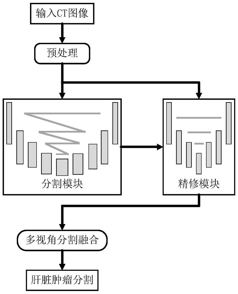
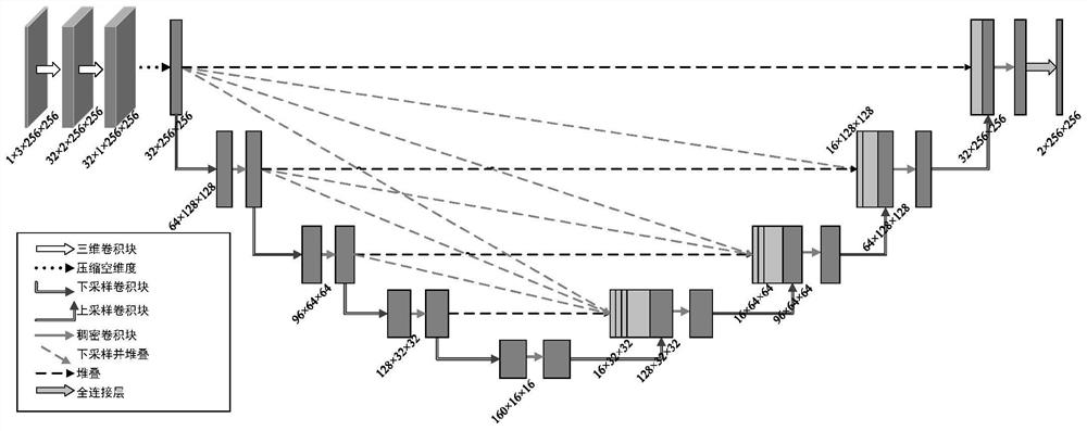
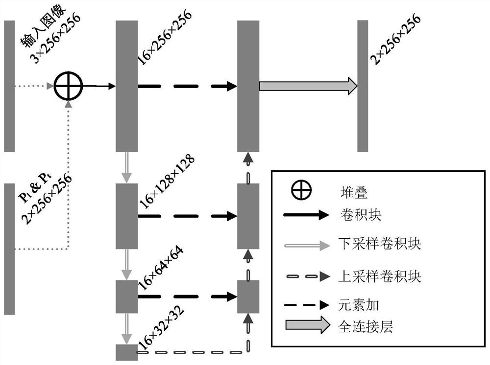
[0045] This embodiment provides a multi-task liver tumor segmentation method based on multi-view. Through this method, the model effectively extracts the three-dimensional space information of the three-dimensional abdominal CT image in a limited graphics card memory environment, and is stable and easy to converge during the optimization process. , to achieve high-precision segmentation of liver and tumor, which can be used in medical image processing.

[0046] The procedure of this method is as follows figure 1 Shown:

[0047] Step 1: Preprocess the 3D abdominal CT image, filter the original image through a threshold to remove pixels outside the gray range of the liver and its tumor, then scale the image size to 256×256×256, and then perform anisotropy on the image Diffusion to reduce the noise in it, and then normalize the image, the remembe...


PUM


Abstract

The invention discloses a multi-view-angle-based multi-task liver tumor image segmentation method. After an abdominal CT image is preprocessed, liver segmentation and tumor segmentation of the abdominal CT image are obtained at the same time in a slice form through a convolutional neural network model. The input of the model is a three-dimensional CT slice with the size of 256 * 256 * 3, and the output of the model is corresponding segmentation of a middle slice. The model comprises a segmentation module and a fine trimming module, and a rough segmentation result and a fine trimming segmentation result are obtained respectively. The model is optimized through a combined loss function, and instability in the optimization process is avoided. According to the method, segmentation is carried out from three perspectives of a three-dimensional CT image, and three segmentation results are fused into one to obtain a final segmentation result. According to the method, liver and tumor segmentation of the abdominal CT image is realized, and the problems that three-dimensional space information cannot be utilized and optimization is unstable in the segmentation process are effectively solved.

Description

technical field [0001] The present application relates to the field of medical image processing, in particular to a multi-task liver tumor image segmentation method based on multi-view. Background technique [0002] Computed Tomography (CT for short) uses precisely collimated X-rays, γ-rays, ultrasound, etc., together with highly sensitive detectors, to conduct continuous cross-sectional scans around a certain part of the human body. , clear image and other characteristics, it can be used for the examination of various diseases. A CT image is composed of a certain number of gray pixels arranged in a matrix, and these pixels reflect the ray absorption coefficient of the corresponding voxel. Based on such medical images, doctors are able to diagnose a patient's condition and assess the patient's response to treatment. [0003] Among them, liver image segmentation using abdominal CT images plays an important role in the field of medical image processing. This is the first st...

Claims


Application Information

Patent Timeline
Patent Type & Authority Applications(China)
IPC IPC(8): G06T7/136G06N3/04G06N3/08
CPCG06T7/136G06N3/08G06T2207/10081G06T2207/30056G06T2207/30096G06T2207/20081G06T2207/20084G06T2207/20221G06N3/045
Inventor 张宇米思娅丁熠玮
Owner SOUTHEAST UNIV
Why Eureka
Try Eureka