Fundus image segmentation model training method and device

A technology for segmenting models and fundus images, which is applied in the field of medical image detection, can solve the problems of affecting model segmentation performance, wasting manpower, and increasing the difficulty of marking objects of interest, so as to improve performance, reduce workload, high practicability and training efficiency Effect

Pending Publication Date: 2020-09-25
SHANGHAI EAGLEVISION MEDICAL TECH CO LTD
View PDF4 Cites 4 Cited by
  • Summary
  • Abstract
  • Description
  • Claims
  • Application Information

AI Technical Summary

Problems solved by technology

To this end, the producer needs to train a high-level model. According to the existing technology, the producer needs to use a large amount of new data to retrain the model. In the new training data, not only the original target of interest needs to be marked, but also the newly

Method used


Image

  • Fundus image segmentation model training method and device
  • Fundus image segmentation model training method and device
  • Fundus image segmentation model training method and device

Examples


Embodiment Construction

[0048] The technical solutions of the present invention will be clearly and completely described below in conjunction with the accompanying drawings. Apparently, the described embodiments are some of the embodiments of the present invention, but not all of them. Based on the embodiments of the present invention, all other embodiments obtained by persons of ordinary skill in the art without making creative efforts belong to the protection scope of the present invention.

[0049] In the description of the present invention, it should be noted that the terms "first" and "second" are used for description purposes only, and should not be understood as indicating or implying relative importance. In addition, the technical features involved in the different embodiments of the present invention described below may be combined with each other as long as there is no conflict with each other.

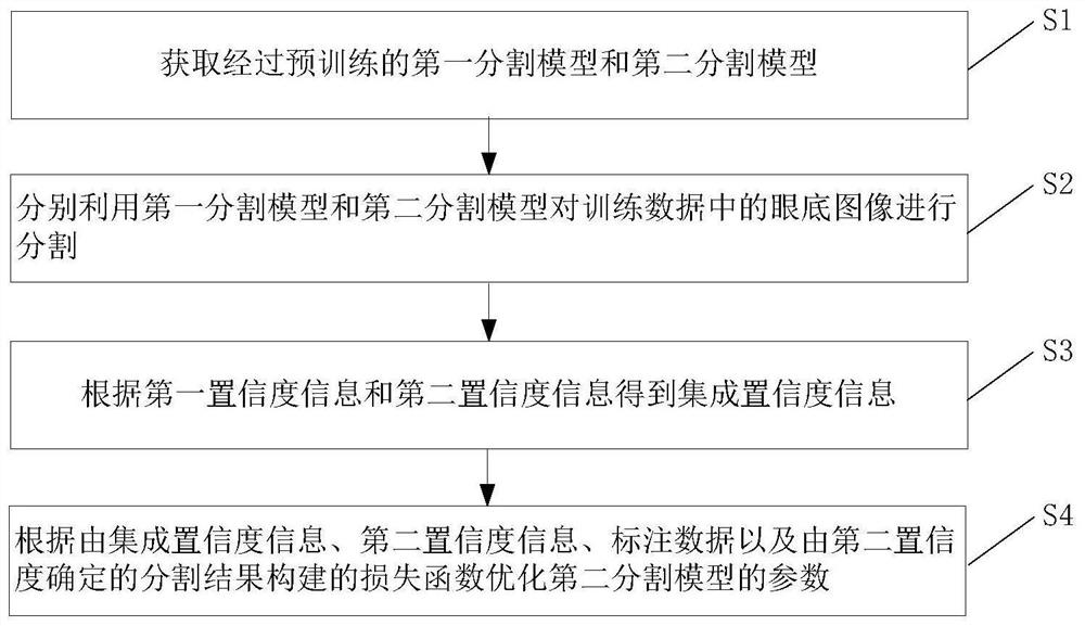
[0050] An embodiment of the present invention provides a method for training a fundus image se...


PUM


Abstract

The invention provides a fundus image segmentation model training method and device. The method comprises the steps of obtaining a first segmentation model and a second segmentation model which are pre-trained; segmenting the fundus image in the training data by using the first segmentation model and the second segmentation model respectively, wherein the first segmentation model outputs first confidence information about the first target of interest, and the second segmentation model outputs second confidence information about the first target of interest and the second target of interest; obtaining integrated confidence information according to the first confidence information and the second confidence information; and optimizing parameters of the second segmentation model according to the integrated confidence information, a second confidence, the annotation data and a loss function constructed by a segmentation result determined by the second confidence.

Description

technical field [0001] The invention relates to the field of medical image detection, in particular to a method and equipment for training a fundus image segmentation model. Background technique [0002] In recent years, machine learning technology has been widely used in the medical field, especially machine learning technology represented by deep learning has attracted widespread attention in the field of medical imaging. In terms of fundus image detection, most semantic segmentation tasks use end-to-end deep learning methods to achieve better results. Accurate boundary and position detection is very important for tracking the development of fundus image lesions, and pixel-by-pixel segmentation of lesions is an important task. A task with great medical application value. [0003] Existing technologies have been able to train machine learning models through a large number of fundus image data sets, so that they can accurately segment objects of interest in fundus images. ...

Claims


Application Information

Patent Timeline
IPC IPC(8): G06T7/194G06T7/11
CPCG06T7/194G06T7/11G06T2207/20081G06T2207/20084G06T2207/30041Y02T10/40
Inventor 贺婉佶熊健皓赵昕和超张大磊
Owner SHANGHAI EAGLEVISION MEDICAL TECH CO LTD
Why Eureka
Try Eureka