Elastin reduction allowing recellularization of cartilage implants

An elastin, recellularization technology, applied in the field of improving cartilage grafts, tissue implants, can solve the problem of not showing cell invasion, etc.

Active Publication Date: 2020-09-25
TRAUMA CARE CONSULT
View PDF11 Cites 1 Cited by
  • Summary
  • Abstract
  • Description
  • Claims
  • Application Information

AI Technical Summary

Problems solved by technology

Furthermore, decellularized natural cartilage implanted under the skin of mice is a highly vascularized environment in which the implants are susceptible to invasion by immune cells, but did not show any signs of cellular invasion (unpublished data)

Method used


Image

  • Elastin reduction allowing recellularization of cartilage implants
  • Elastin reduction allowing recellularization of cartilage implants
  • Elastin reduction allowing recellularization of cartilage implants

Examples


Embodiment 1

[0083] Example 1: Recellularization with BMSCs by dynamic seeding

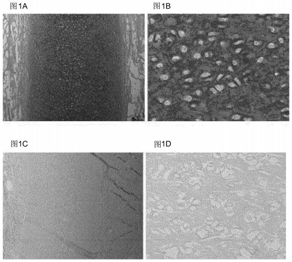
[0084] Wash the outer ear of a 12-week-old calf with water, and remove the perichondrium, skin, and muscle with a scalpel until only cartilage remains. Subsequently, biopsy samples with a diameter of 6 mm and a height of approximately 1 mm are obtained from the ear cartilage using a biopsy punch, so each sample has a volume of approximately 30 mm 3 .

[0085] Cells and elastin fibers were then removed using the following protocol, volumes given are for batches of approximately 20 samples unless otherwise stated. First, the samples were briefly rinsed three times with 40 ml. In some embodiments of the invention, 3696 Grade I deionized water (hereafter referred to as deionized water) is used to remove debris, such as loose pieces of skin, from the isolate. If necessary, samples were stored frozen in 20 ml of deionized water at -20°C. The samples were then sterilized with 20 ml of 5% hydrogen peroxide for 60 mi...

Embodiment 2

[0089] Example 2: In situ cartilage formation after injection

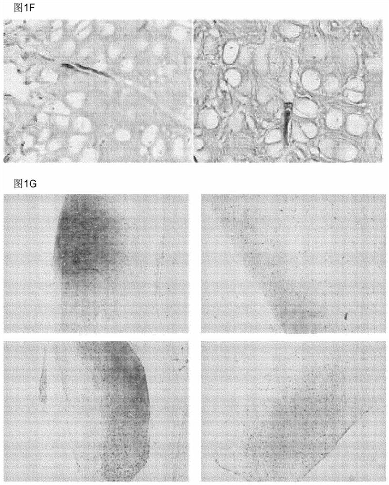
[0090] Certain areas of cartilage have difficulty reaching cells by migration, for example, the cartilage of the outer ear is surrounded by a dense layer of perichondrium. As shown by Utomo et al. 2015, whole ears can be decellularized using elastase. The perichondrium is tightly attached to the cartilage, and it is not feasible to remove it without damaging the cartilage structure.

[0091] To test whether recellularization of the whole ear is feasible, bovine ear cartilage samples were recellularized with chondrogenic cells by perichondrial injection without having to remove the perichondrium.

[0092] The external ear of a 12-week-old calf was washed with water, and the skin and muscle were removed with a scalpel until only the cartilage of the intact perichondrium remained. Subsequently, square biopsy samples with a side length of 15 mm are obtained from the ear cartilage using a scalpel, and each sample has...

Embodiment 3

[0097] Example 3: In vivo cartilage formation of joint models

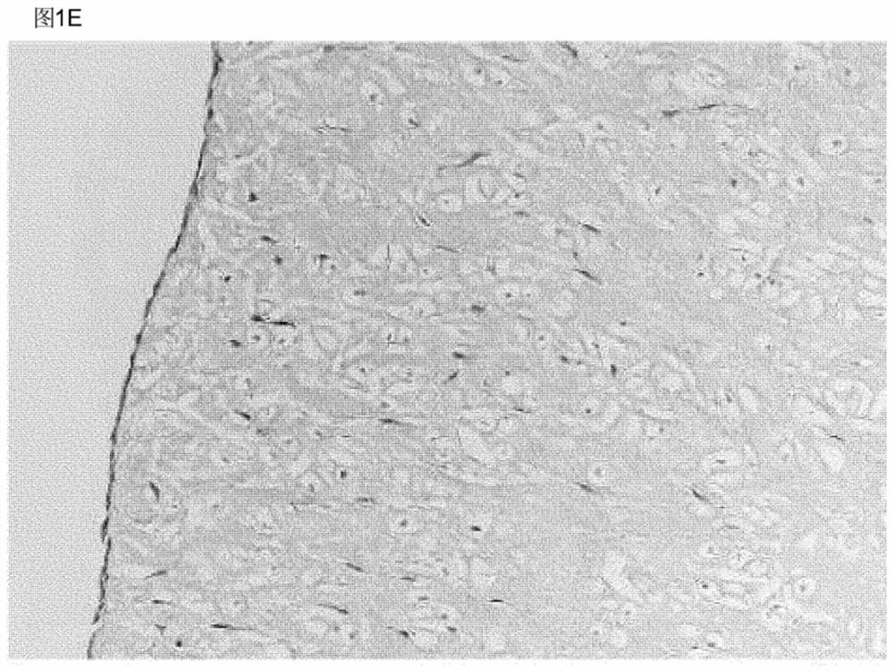
[0098] To determine whether elastase-treated elastic cartilage is suitable for repairing cartilage defects, it was used to fill defects in an articular cartilage injury model. First, the outer ear of a 12-week-old calf was washed with water, and the perichondrium, skin, and muscle were removed with a scalpel until only cartilage remained.

[0099] Subsequently, a 6 mm diameter biopsy was obtained from the ear cartilage using a biopsy punch and cut to a height of approximately 0.4 or 1.2 mm.

[0100] The protocol used to remove cells and elastin fibers was the same as that described in Example 1. Volumes given are for batches of approximately 20 samples unless otherwise stated.

[0101] After removing cells and elastin fibers, place half of the sample in a 0.3 cm 2The bottom of the well (one sample / well, 96-well plate), and cover 500,000 chondrocytes in each sample, as a solution, add to 200 μl of chondrocyte ex...


PUM

PropertyMeasurementUnit
thicknessaaaaaaaaaa

Abstract

The invention relates to a method of producing an elastin-reduced cartilage scaffold containing channels and / or lacunae, the method comprising the steps of providing an elastic cartilage sample and reducing elastin from said cartilage sample to produce said channels and / or lacunae. The invention further relates to an elastin-reduced cartilage scaffold. Said elastin-reduced cartilage scaffold can be used in a method of implantation in vivo, for repairment of cartilage, repairment of osteochondral defects and repairment of bone defects.

Description

technical field [0001] The present invention relates to the field of tissue implants. More specifically, the invention is in the field of improved cartilage grafts. In particular, the present invention relates to a method for reducing the elastin of cartilage scaffolds containing channels and / or lacunas allowing recellularization of the scaffold. Background technique [0002] Bodily organs may decline due to congenital disorders, acquired disease, accidents and old age. The scarcity of donor organs and the need to suppress the immune system to prevent organ rejection both make organ transplants difficult. Over the past few decades, our knowledge of the cells that make up organs has expanded rapidly, making the creation of transplanted organs from scratch (so-called tissue engineering) increasingly realistic. An organ is a combination of cells and a matrix, the latter providing the 3-dimensional structure that holds the cells in place. The cells carry markers unique to ea...

Claims


Application Information

Patent Timeline
Patent Type & Authority Applications(China)
IPC IPC(8): A61L27/36A61L27/38A61L27/56
CPCA61L27/3612A61L27/3687A61L27/3817A61L27/3834A61L27/56A61L2430/06A61L2430/40A61L27/3654A61F2/30756A61F2002/30766A61F2002/30762A61L2430/02A61L27/3691
Inventor G·J·V·M·范·奥施J·莱曼S·尼恩贝格尔
Owner TRAUMA CARE CONSULT
Why Eureka
Try Eureka