

Animal tissue transparentizing method
An animal tissue and tissue technology, applied in the field of animal tissue observation, can solve the problems of protein observation, difficulty in imaging, tissue deformation, etc., and achieve the effects of shortened transparency time, high safety, and easy operation.
- Summary
- Abstract
- Description
- Claims
- Application Information
AI Technical Summary
Problems solved by technology
Method used
Image

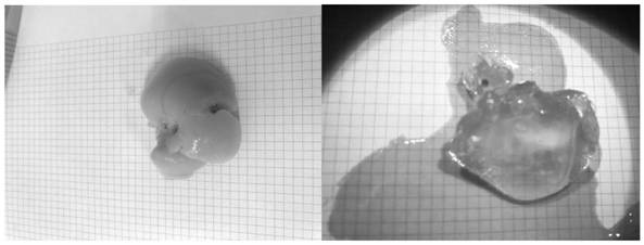
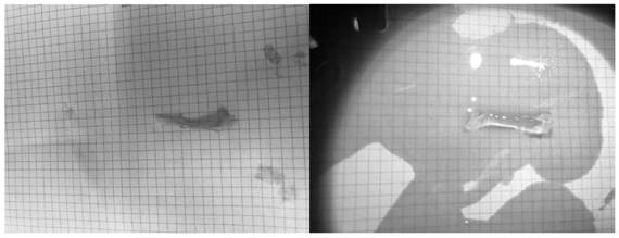
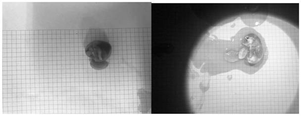
Examples
Embodiment 1
[0047] Reagents required for the experiment:
[0048] Anesthetic: 0.3% pentobarbital (0.3g pentobarbital dissolved in 100mL deionized water) and ketamine (90mg / kg);
[0049] Phosphate buffered saline solution (PBS): 1×PBS is 0.01MPBS, after dissolving 22.4g (phosphate buffered solution, PBS) buffer dry powder with 2L deionized water, adjust the pH value to 7.2-7.4.
[0050] Tissue fixation solution: 4% paraformaldehyde, that is, 4g of PFA (Paraformaldehyde, PFA) powder dissolved in 1×PBS to 100mL;
[0051] Cardiac perfusate: 0.9% normal saline injection or 0.05 MEDTA, that is, 0.05 mol ethylenediaminetetraacetic acid (EDTA) is dissolved in deionized aqueous solution to a total volume of 1 L;
[0052] 0.1% Triton solution: Put TritonX-100 in a water bath at 37°C to 40°C for 2 to 3 hours to fully dissolve and mix well, then take 100μL TritonX-100 and dissolve it in 1×PBS to 100mL.
[0053] 3% BSA solution: Dissolve 3g of BSA powder in 0.1% Triton solution to 100mL.
[0054] 5...
PUM
 Abstract
Description
Claims
Application Information